
AMBULATORY MONITORING OF QKD TO ASSESS ARTERIAL DISTENSIBILITY
Philippe GOSSE MD
Hopital Saint André
BORDEAUX. France
E-mail: philippe.gosse@chu-bordeaux.fr
| DISCUSSION BOARD |
INTRODUCTION
1. Introduction
The physical properties of the large arteries and, in particular, the aorta, play an important role in cardiovascular physiology and physiopathology. Reduced aortic distensibility is certainly a contributing factor in mortality and morbidity linked with age and arterial hypertension 1-3 and very probably in other pathological circumstances too (diabetes, systemic sclerosis, etc.). The oldest technique for assessing these physical properties is measurement of the PWV. The principle of this technique is fairly simple: the more rigid the artery, the faster the vibration generated by the ventricular ejection is transmitted by the arterial wall throughout the whole arterial tree, which is the origin of the pulse. Although the principle is simple, this method bas several drawbacks, which have limited its use in everyday clinical practice. More precise and sophisticated new techniques4 are now available, based on instantaneous measurement of pressure and of the arterial diameter, enabling the Diameter/pressure curve and arterial compliance to be calculated during the systolic-diastolic cycle (dD/dP). These methods are, however, restricted to the superficial arteries (whose properties may be different from those of the aorta) and, above all, the equipment is expansive and the results depend on the operator's skill, and so only a few teams are able to use this technique. It remains difficult, therefore, for a clinician today to be able to assess the physical properties of the large arteries, apart from simple arterial pulse pressure measurement, whose elevation is often, but not exclusively, due to reduced aortic distensibility (increased cardiac output, aortic regurgitation, etc.).
We have developed a new technique, based on ambulatory measurement of the timing of the appearance of Korotkoff sounds, which enables the variations of a parameter reflecting the PWV to be measured according to the variations in blood pressure. This method provides indications about the arterial rigidity automatically during a 24 hour period of ambulatory blood pressure monitoring. Its main advantages are that it is automatic, simple to use, and is combined with the benefits of ambulatory blood pressure measurement. This technique therefore allows clinicians in their everyday practice to assess the physical properties of the large arteries.
2. Physical properties of the large arteries - consequences
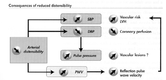
The elastic fibres in the arterial wall and, in particular in the aorta, buffer the systolic wave, enabling the discontinuous flow (systolic) generated by the left ventricle to be transformed into a continuous flow (systolic-diastolic), which is much more efficient in terms of output. Reduction of aortic distensibility or compliance has several harmful consequences . Reduction of the buffering function results in an elevation of the systolic pressure, reinforced by the precocious return of the reflection waves due to increased PWV, and a fall in diastolic pressure. The elevation of the systolic pressure increases the cardiac work during ventricular ejection and increases the risk of development of left ventricular hypertrophy (LVH). At the vascular level, elevation of the systolic BP is a risk factor for strokes. Reduced diastolic BP may impair the coronary perfusion pressure and if it is too low it can be particularly harmful for a patient with LVH if the coronaries have lost part of their vasodilatation properties, through the effects of hypertension.
Therefore, alteration of aortic distensibility certainly contributes indirectly to the well recognised risk associated with LVH. Finally, elevation of the pulse pressure may, in itself, constitute a factor of mechanical alteration of the arterial wall and contribute with heart rate to increasing the rate of cardiovascular complications5 . Elevation of the pulse pressure is also an important stimulus to the development of hypertrophy of the media at the level of the muscular arteries, leading to increased peripheral resistance. Arterial pulse pressure appears to be a risk factor independent from the mean BP6-7 , which is, in all probability, the result of the damaging effects of alteration of the aortic distensibility. However, if pulse pressure measurement can be considered as a first simple approach to this phenomena, it remains non specific. Specific means of exploring the physical characteristics of the large arteries are indispensable for better assessment of the alteration of these properties in a certain number of pathologies as hypertension, their consequences and the effects of therapeutic treatment.
3. Pulse wave velocity measurement
PWV measurement is the oldest method available for estimating the rigidity of an arterial segment8. The more rigid the arterial segment is, the more rapidly the systolic pulse, will travel the arterial wall. Despite the length of time it bas been available and the simplicity of its concept, this method bas remained little used for various reasons:
4. QKD interval
4.1 Definition
The QKD interval corresponds to the time which separates the QRS complex from the appearance of the last Korotkoff sound detected by a microphone placed over the brachial artery during the BP measurement; it corresponds therefore to the moment when diastolic BP is detected. Therefore Q stands for QRS, K for Korotkoff and D for diastolic (fig.2). This interval has a duration of about 210 milliseconds in young subjects at rest11.
This time can be divided into the sum of 2 phenomena:
a)The pre-ejection time which starts with the QRS complex and ends with the beginning of the left ventricle ejection phase (opening of the aortic valves). The pre-ejection time is itself made up of 2 components:
The pre-ejection has a mean duration of 70 to 110 milliseconds in normal subjects at rest.
b)The pulse wave transmission time on a territory comprised between the aortic valves and the microphone placed over the brachial artery which includes therefore the ascending aorta, the sub-clavian artery and the main part of the brachial artery. This transmission time therefore depends on the length of this arterial segment and the PWV. Any elevation of the PWV will lead to a shortening of the pulse wave transmission time and therefore of the QKD interval. This technique is therefore very close to measurement of the PWV and has 2 major advantages: measurement of the parameter is completely automatic and it can be measured for different BP levels. It is the variations of this time according to the BP which make this technique valuable, the effect of the pre-ejection time having to be taken into account, or restricted.
4.2 Historical background
To the best of our knowledge, the first person to study the timing of the appearance of Korotkoff sounds was Simon Rodbard12 . Dr Rodbard has published several studies dedicated to the measurement at rest of the interval separating the QRS complex of the ECG from detection of the arterial sounds at the level of the brachial artery, during cuff deflation, between the systolic BP (appearance of the first sound, QKS) and the diastolic BP (last sound, QKD). His idea was that the progressive decrease of this interval during cuff deflation follows a curve reproducing the shape of the intra-arterial wave. This coincidence in shapes led his studies, in our opinion, in the wrong direction, but he was, however, able to describe different factors influencing the variations of these intervals. In particular, it showed that the timing of the appearance of arterial sounds shortens with effort. Later on Dr Rodbard and others who continued these experiments, studied this timing in particular in relation to the preejection time, as an indication of the systolic function. The QKD interval was proposed as a means of monitoring dysthyroïdism13 : it diminishes in cases of hyperthyroidism and rises in cases of hypothyroidism. More recently14, however, the interest of using the QKD interval as an indication of the PWV was put forward, but without any practical follow-up.
We rediscovered this interval while developing automatic, auscultatory devices for the measurement of blood pressure during exercise. Systematic recording of the timing of the onset of arterial sounds in relation to the ECG at rest and during exercise led us to study this parameter as a possible reflection of PWV. These studies, which started in 1988 with an ECG recording device modified to incorporate the signal of the microphone placed on the brachial artery, and continued with a prototype designed by NOVACOR France for automatic measurement of the QKD interval, have enabled us to confirm the relevance of the QKD interval. In 1991, this measurement was integrated into an ambulatory BP monitor
4.3 Automatic measurement of the QKD interval at rest
The QKD interval is measured automatically during the BP measurement between the deflection of the QRS complex and the onset of the last Korotkofff sound with an accuracy of 5 milliseconds. The cuff is usually placed on the left arm with the microphone over the brachial artery. Three electrodes are placed on the chest for detection of the ECG and the measurement is taken with the patient lying down or sitting. The measurement at rest is the mean of at least three successive measurements of the BP and the QKD
The reproducibility of this measurement was studied in 31 patients - 17 normotensives and 14 hypertensives - by repeating 5 measurements at rest at a fortnight's interval. The mean value was from 205 ±23 milliseconds the first day and 206 ±24 milliseconds a fortnight later. The mean of the differences is 1 ±15 milliseconds, i.e. a coefficient of variation of 7.3%.
The correlation between the QKD interval and the PWV was studied in 37 subjects - 20 normotensive and 17 hypertensive11. The PWV was measured between the subclavian and the radial arteries. The pressure wave was detected by continuous Doppler recording between these points. The recordings were made at a speed of 100 mm/s. Correlation with the QKD interval measured simultaneously on the other arm was significant, but weak (r=0.55, p<0.01). Significant but weak er correlations between QKD and PWV on descending aorta at rest were also found by Abassade15 in 62 patients. There are several reasons for this poor correlation: the difference between the territories considered by the 2 methods, a lack of reliability of the PWV measurement, and finally above all, the influence of the pre-ejection time on the QKD measured at rest. It is therefore vital for the use of the QKD as an index of arterial rigidity to eliminate the effect of the pre-ejection time. This can be achieved with ambulatory measurement of the QKD combined with measurement of the BP and heart rate, as we will explain later on.
4.4 Factors of variations of the QKD interval
4.4.1 Influence of cuff pressure
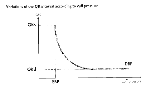
The timing of the appearance of Korotkoff sounds (QK) diminishes progressively during cuff deflation. The QK interval reaches its maximum value during the appearance of the first arterial sound (QKS=320 ±20 milliseconds) and falls progressively with cuff deflation in a curvilinear way. The shape of this curve looks very similar to the decrease of the pressure wave in the artery. In fact, the transmission time of arterial sounds at the level of the microphone in the cuff is slowed down because the cuff is compressing the artery. This effect is reduced with cuff pressure and disappears several mm of mercury above the diastolic pressure, which is why there is a plateau in the QK interval at the diastolic level . Only this measurement of the QKD has a physiological significance. This relationship between the cuff pressure and the delay before the appearance of arterial sounds enables an important source of artefacts in the automatic measurement of the QKD to be apprehended. When detection of arterial sounds is poor, the device may stop listening before the diastolic pressure is actually reached. The result is an over-estimation of the diastolic BP and the QKD interval which is measured on the descending portion of the curve and not on the plateau. This is the reason why the study of the relationship between the QKD and the diastolic BP is so important in ambulatory recordings, as we will show later on
4.4.2 Influence of arterial Pressure
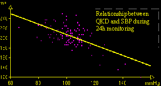
The QKD interval falls when arterial pressure rises. This is essentially the reflection of the relationship between PWV and BP. The relationship between PWV and BP was studied in rats in the laboratory of Professor Freslon in Bordeaux16. using miniature intra-arterial sensors placed on the aortic arch, over the left carotid and in the abdominal aorta over the iliac bifurcation. The distance between the two sensors was measured accurately with an X-ray photo. The shift of the pressure wave between the two sensors was measured on the simultaneous and amplified recording of pressure signals. The animals' BP was modified by Noradrenaline or sodium nitroprussiate injections. The BP and the PWV varied in the same direction. The relationship between the two appeared to be linear for both experiments (noradrénaline and niitroprussiate) which examined a fairly limited pressure field. However, in the same rat, the combination of points obtained with the 2 drugs enabled a much larger pressure field to be examined and a curvilinear relationship to be noticed. The curvilinear character of this relationship was expected. It corresponds to the curvilinear character of the relationship between diameter and pressure or the relationship between compliance and pressure of the artery which has been well established in vitro and in vivo, both in man and in animals. But, in order to prove the curvilinear character, examination of a wide range of pressure is necessary. The QKD and blood pressure are inversely related. When the BP rises, the QKD interval falls. This relationship is shown in individuals, for example during exercise and particularly during spontaneous BP variations of the pressure during ambulatory monitoring of the BP and the QKD interval. There is a strong correlation between BP variations, whether recorded at test or during exercise, and the QKD interval. This correlation is very significant with the systolic BP, and often even more so with the pulse pressure. Correlation with diastolic BP is, in general, much weaker and often not significant . The relationship between the QKD and systolic or pulse pressure, is, in general, linear. This is the same as in animals, no doubt due to examination of too narrow a pressure range for the curvilinearity of the relationship to appear. However, we were able to show the curvilinear character of this relationship by superimposing recordings taken in hypertensive patients before and shortly after the start of an antihypertensive treatment inducing an important lowering of BP.
4.4.3 Influence of the heart rate
The QKD interval is correlated negatively to the heart rate. This correlation is, in general, weaker than the correlation observed with the BP. It is particularly clear during physical exercise whereas it is not always obvious during spontaneous variations of the BP and the heart rate. This relationship to the heart rate is linked to the influence of the pre-ejection period (PEP) on the QKD interval. When the QKD interval is used as a reflection of PWV, the effect of the pre-ejection period, which depends on inotropism, is a undesirable factor. The relationship between the PEP and the heart rate is a linear relationship in which the variation depends on a sympathetic stimulation of the system, such as during exercise. This relationship has been known for a long time and Weissler17 proposed an adjustment formula of the PEP according to the heart rate (adjusted PEP (ms) = PEP + 0.4 HR) from studying variations of the PEP according to the spontaneous variations of the heart rate. Murata and colleagues18 found a negative correlation between QKD and heart rate in a normal population with different slopes according to sex:
Males: n=78, r=-O.24, p<0.05, QKD=-O.469 HR +247
Females: n=47, r=-O.46, p<0.01, QKD=-0.799 HR +270
We tried to use Weissler's, Murata's and other equivalent formulas obtained in our normal population, to adjust the QKD interval. This type of correction always resulted in increasing the link between the heart rate and adjusted QKD as opposed to the non-adjusted QKD, which underlined it is inadequate. In fact, the relationship between the QKD and the heart rate depends on the individual and the conditions of the heart rate variations. In rats, a linear relationship between the PEP and heart rate was found when dobutamine was injected, with large variations in slopes between one animal and another16 . In man, the relationship between the QKD and the heart rate also varies greatly from one individual to another and, no doubt, from one moment to another. It therefore seems better to define this relationship for each individual and for each recording. It can then be used to limit the consequences of the PEP variations on the interpretation of the QKD variations.
4.4.4 Influence of height
The length of the arterial segment along which the pulse wave travels has, of course, an effect on the pulse wave transmission time and therefore on the QKD interval. ln a study carried out on children and adolescents between 7 months and 18 years old, Bercu and colleagues14 showed that it was of no use to try to measure the distance between the aortic valves and the cuff microphone and that the best way of correcting the QKD interval in order to make comparisons between subjects, was by taking the subject's height into consideration. In this population, the relationship between the QKD measured at rest and the subject's height was:
QKD (ms) = 0.797 height (cm) + 56.63.
In a population of normotensive adults we found a significant, but poor correlation (n= 1 15, r=O. 17, p<O.O 1) between the QKD measured at rest and height: QKD (ms) = 0.43 height (cm) + 127. We proposed that this factor be taken into account by expressing the QKD measured as a percentage of the value predicted by the subject's height.
4.4.5 Influence of age
The QKD interval measured at rest falls significantly with age and this can only be linked with the rise in the PWV, illustrating the well established increase of arterial rigidity with ageing. This can only be seen in adults because an inverse relationship is observed in children due to the predominant effects of growth, whereas the PWV is not altered14. Tables 1 and 2 give the normal values of the QKD interval according to age in Murata's population18 and in ours. These values, although recorded in very different populations with very different devices, were very similar. A reduced QKD interval is obvious after 60 years of age in normal subjects. In our normal population there was a significant negative correlation between the QKD measured at rest and age. This has also been found recently by Bulpitt19 who have reported that among different measures of vascular compliance the QKD interval shows the best correlation to age.
Values of the QKD interval measured at rest in a normal population (Murata, 1976)
|
Age (years) |
6-9 |
10-20 |
20-30 |
30-40 |
40-50 |
50-60 |
60-70 |
>70 |
|
MALES |
|
|
|
|
|
|
|
|
|
Number |
5 |
19 |
48 |
19 |
11 |
10 |
7 |
13 |
|
QKD (ms) |
171±10 |
196±18 |
221±14 |
211±14 |
209±14 |
192±10 |
193±20 |
185±14 |
|
FEMALES |
|
|
|
|
|
|
|
|
|
Number |
5 |
13 |
23 |
13 |
11 |
10 |
6 |
11 |
|
QKD (ms) |
161±10 |
186±16 |
214±17 |
209±10 |
213±20 |
208±27 |
191±14 |
179±14 |
Values of the QKD interval measured at rest in a normal population (Gosse, 1991)
|
Age (years) |
20-30 |
30-40 |
40-50 |
50-60 |
>60 |
|
Number |
33 |
37 |
20 |
12 |
8 |
|
Height (cm) |
170±8 |
168±6 |
166±9 |
167±9 |
162±8 |
|
SBP (mmhg) |
123±14 |
121±11 |
122±14 |
126±13 |
133±15 |
|
DBP (mmhg) |
77±10 |
76±11 |
80+12 |
84±10 |
86+7 |
|
HR (bpm) |
74±15 |
77±13 |
71±15 |
71±8 |
73±13 |
|
QKD (millisec) |
206±17 |
198±17 |
202±15 |
198±9 |
178+11 |
4.4.6 QKD and conduction problems
The pre-ejection period is lengthened in patients with left bundle branch block (electromechanical interval). In this case the QKD interval is also lengthened. It would, no doubt, be possible to adjust the QKD by taking into account the width of the QRS complexes, but we have not verified this yet.
4.4.7 Other factors
In our experience, the arm used for the QKD measurement is of no importance. We have not found any systematic difference between men and women either, as long as the differences in height and BP are taken into account.
5. Ambulatory measurement of the QKD Interval
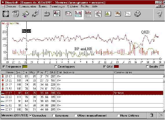
Ambulatory measurement of the QKD interval enables the variations of this interval in relation to the spontaneous variations in BP and heart rate to be studied. It also enables to assess the relationship between PWV and BP and to limit the influence of the variations in pre-ejection time by studying the relationship between the QKD and heart rate20.
5.1 Method
The QKD interval is measured every 15 minutes over a period of 24 hours, at the same time as the BP and the heart rate, with an ambulatory measurement device using the auscultatory method with 3 electrodes for detection of the ECG signal (DIASYS INTEGRA, NOVACOR, Rueil-Malmaison, France http://www.novacor.fr/. The data can now be interpreted automatically with the DiasySoft software (NOVACOR, France). This software displays the BP, heart rate and QKD measurements in a table and a graph. Any abnormal values beyond the usual thresholds of interval variation (100-300 ms) are automatically eliminated. An assessment of the whole set of values, made easier by examining the graph, enables any other abnormal values, which may occur if the measurement was taken in difficult conditions, to be removed manually by the clinician. The program then displays the variations of the QKD interval in relation to the systolic BP, diastolic BP, pulse pressure and heart rate in graphs, and gives the linear equation linking the QKD to each of these parameters.
There is usually a very clear negative relationship between the QKD and the systolic blood pressure or pulse pressure which illustrates the relationship between the PWV and BP. The relationship with the diastolic blood pressure is generally weaker. A strong positive correlation between the QKD interval and the diastolic BP usually means that there has been a problem in recording. This usually corresponds to an overestimation of the diastolic blood pressure and proportionally to the QKD interval following faulty detection of the Korotkoff sounds. This type of recording should therefore be deleted. However, it should be noted that a positive relationship between the diastolic blood pressure and the QKD can also be observed in young subjects performing physical exercise during the recording. In fact, in young subjects exercising a fall in diastolic pressure is observed, whereas the systolic BP rises and the QKD falls. These graphs therefore enable the quality of the recording to be checked. The program also calculates the QKD100-60 This index is one of the key elements of the interpretation. It corresponds to the value of the QKD interval for a systolic BP of 100 mmhg and a heart rate of 60 bpm. It is calculated from the following equation: QKD =C - ASBP - bHR obtained from the measurements of the recordings. This normalised QKD enables inter- and intra-subject comparisons to be made for different BP levels and the variation of the QKD linked to the heart rate and therefore to the pre-ejection period, to be eliminated21.
Up to now, our studies have been based on 24 hour recordings. This duration bas two advantages: first of all it means that, by taking 4 measurements an bout, a large number of measurements (about one hundred) are obtained, enabling the relationships between the QKD and BP and the QKD and heart rate to be defined, and secondly, by including both night-time rest and daytime activity, it enables to assess a fairly wide range of spontaneous variations between BP and QKD . It is however possible to use recordings taken over a shorter period of time with standardised conditions concerning BP variations (rest period, standardised physical activity)22.
5.2 Interpretation
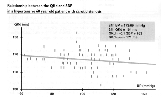
Increase in the stiffness of the arterial system results firstly in a fall in the QKD interval and in particular its mean value over 24 hours, because of the increase of PWV, and secondly in a decrease in the effect of BP variation on PWV and therefore the QKD interval. This can be explained by using a simple image. In a flexible tube, the transmission speed of parietal vibration will depend on the distension of the wall by the internal pressure of the liquid, but, by contrast a very rigid metallic tube will stay just as rigid whatever the internal pressure is. In the same way the increase in arterial rigidity will lead to a decrease in the slope linking the QKD interval to the systolic or pulse pressure and to a reduction of the QKD100-60. Figure illustrates the result of an ambulatory measurement of the QKD interval in a hypertensive 60 year old patient with carotid stenosis. The mean QKD interval over 24 hours is reduced to 164 ms, the variation slope of the QKD interval according to the systolic BP is almost zero, the QKD100-60 bas a reduced value: 171 ms.
5.3 Reproducibility
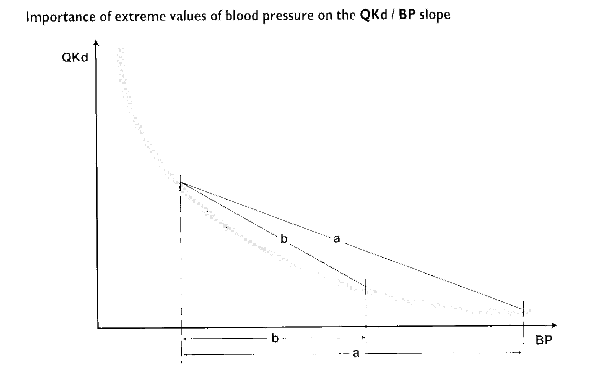
Reproducibility was studied in 28 normal subjects - 14 men and 14 women aged from 26 to 77 (average 43 ±16 years) - who had two 24 hours recordings taken during their usual activities, separated by one week23. The table below summarises the main results. There is no relationship between the importance of these differences and the value of the parameter. The reproducibility of the mean QKD over 24 hours and of the QKD100-60 seems good, at least as good as the reproducibility of the mean BP levels. This is not the case for the variation slopes of the QKD according to the BP. These are not very reproducible. This is linked to the tact that the relationship between the QKD and the BP is curvilinear rather than linear. Thus the slope calculated on a linear relationship strongly depends on extreme values, as illustrated in figure. These extreme values are not very reproducible from one procedure to another, although the mean values are. A way of improving the slope reproducibility could be to get the subject to perform a standardised physical effort during the recording which would enable a maximum reproducible value of the BP to be obtained (and a minimum of the QKD).
Reproducibility of the main parameters provided by ambulatory measurement of the QKD interval
|
|
1rst |
2nd |
SDD |
CV |
r |
|
24h SBP(mmHg) |
116±22 |
113±18 |
9 |
8% |
0.91 |
|
24h DBP (mmhg) |
74±9 |
73±7 |
5 |
5% |
0.84 |
|
24h HR (bpm) |
77±9 |
76±10 |
5 |
60/. |
0.86 |
|
24h QKD(ms) |
190±20 |
190+19 |
7 |
5% |
0.90 |
|
QKD100-60 (ms) |
200±21 |
199+17 |
1.2 |
6% |
0.83 |
|
Slope/SBP |
0.41±0.24 |
0.44±0.31 |
0.31 |
49% |
0.29 |
|
Slope/pulse pressure |
0.75±0.33 |
0.75±0.31 |
0.32 |
42% |
0.52 |
5.4 Influence of height
Like the QKD at rest, the QKD100-60 depends on the subject's height. This relationship was studied24 in a population of 85 normal subjects less than 30 years old with no known factors of risk (smoking, cholesterol). Correlation between the QKD100-60 and height is significant: : r = 0.50, p < 0.02, QKD100-60 = 91 + 0.73 x Height. This equation enables the theoretical value of each individual's QKDIOO-60 to be calculated and the QKD100-60 observed to be expressed as a percentage of this theoretical value. This calculation is automatically made by the DiasySoft Integra software (NOVACOR, France). This percentage is no more correlated to height.
5.5 Variations according, to age
With age there is a significant reduction of the mean QKD over 24 hours, of the slopes according to the BP and of the QKD100-60. This illustrates the increase of arterial stiffness with ageing. This relationship was studied in a population of 180 normal subjects24. The relationship between the QKD100-60 and age is very significant and independent from the mean level of the ambulatory BP.
5.6 Variations in hypertensive subjects
5.6.1 Comparison between hypertensives and normotensives matched by age
We compared untreated subjects with essential hypertension with normal subjects, matched by age (older or younger than 40). The main results are shown in table:
Comparisons between essential hypertensives and normotensives matched by age
|
|
< 40 years |
>40 years |
||
|
|
Normotensives |
Hypertensives |
Normotensives |
Hypertensives |
|
Number |
97 |
28 |
60 |
95 |
|
Age (years) |
27±8 |
30±6 |
51±9 |
55±10 |
|
24h SBP (mmhg) |
110±10 |
132±15 |
109±9 |
135±17 |
|
24h DBP (mmhg)70±9 |
86±16 |
75±6 |
87±11 |
|
|
24h HR (bpm) |
77±10 |
83±10 |
74±10 |
77±9 |
|
24h QKD (ms) |
204±14 |
196±13 |
193±16 |
183+17 |
|
QKD100-60 (ms) |
214±15 |
216±17 |
200±17 |
194±20 |
|
Theoretical QKD |
1±0.06 |
1±0.08 |
0.95±0.08 |
0.91±0.09 |
|
SBP slope |
-0.70±0.34 |
-0.58±0.28 |
-0.50±0.32 |
-0.36±0.21 |
|
Pulse pressure slope |
-1.03±0.41 |
-0.76±0.19 |
-0.93±0.32 |
-0.63±0.31 |
The slope of the QKD against the systolic BP or the pulse pressure is significantly reduced in hypertensive subjects in both age groups. In contrast the QKD100-60, as an absolute value or as a percentage of the theoretical value, is only significantly reduced in hypertensives over 40 years old.
A multivariate analysis shows that the age and the mean systolic BP over 24 hours are both significantly (p<0.001) and independently correlated to the QKD100-60, in the whole population as well as in hypertensive patients.
5.6.2 relationship to left ventricular mass
We found a very significant correlation between different parameters resulting from the ambulatory measurement of the QKD and left ventricular mass or relative wall thickness25 measured by echocardiography in a population of untreated hypertensive subjects. This relationship underlines the role of alterations in the physical properties of the large arteries in the development of a left ventricular hypertrophy in hypertensives. This role has already been underlined with other measurement techniques: PWV26, and invasive measurement of aortic impedance27.
5.7 Reference values
5.7.1 Reference values by age group
Table illustrates the values of the different QKD parameters obtained by ambulatory measurement over 24 hours by age groups in a population of 157 normal subjects.
Values of the parameters obtained in a normal population according to age
|
Age (years) |
10-20 |
20-30 |
30-40 |
40-50 |
50-60 |
>60 |
|
Number |
1 5 |
48 |
28 |
37 |
20 |
9 |
|
Weight (Kg) |
65±1 0 |
66±14 |
68±13 |
70±13 |
72±10 |
64±11 |
|
Height (cm) |
173±10 |
172±9 |
170±9 |
169±9 |
168±9 |
162±7 |
|
24h SBP (mmhg) |
109±9 |
110±11 |
109±8 |
107+9 |
112±8 |
108±8 |
|
24h DBP (mmhg) |
65±7 |
69±9 |
74±6 |
76±7 |
76±6 |
72+3 |
|
24h HR (bpm) |
75±11 |
76±11 |
77+9 |
77±10 |
73±10 |
71±9 |
|
24h QKD (ms) |
204±13 |
208±15 |
198±13 |
199±14 |
194±16 |
178±19 |
|
QKD100-60 (ms) |
215±15 |
217+15 |
208±11 |
206±16 |
203+17 |
186+16 |
|
QKD100-60 % |
100±7 |
101±6 |
97±6 |
97±7 |
96±10 |
89±6 |
|
Slope/SBP |
0.90±0.38 |
0.74±0.29 |
0.54±0.31 |
0.56+0.40 |
0.46±0.22 |
0.51±0.24 |
|
Slope/PP |
1.17+0.49 |
0.97±0.39 |
1.04±0.38 |
1.03±0.36 |
0.84±0.24 |
0.78±0.28 |
These subjects were volunteers, some of whom were recruited in the medical environment, but most were recruited at their workplaces in a company in Bordeaux. These subjects had never been treated and had no history of cardiovascular diseases. The results are shown in 10 year age groups. All had normal BP measured over 24 hours. The systolic BP is not significantly different between the age groups. The diastolic BP rises with age in the first 5 decades and tends to fall after 60 years of age. There is a significant correlation with age for all the QKD parameters studied. The QKD100-60 and its percentage value of the theoretical value predicted by height are significantly negatively correlated to age and to mean systolic BP level, both contributing significantly to the relationship in a multivariate analysis.
5.7.2 Normal value of the QKDIOO-60 interval
This normal value was calculated in a population of 40 normal subjects under 40 years old and without any risk factor. The mean value of the QKD100-60 in this population is 214 ±14 ms with values of 187 to 242 ms. The lower limit of the normal value defined as the mean less 1.96 standard deviation is 187 ms. When the QKDi100-60 is expressed as a percentage of the theoretical value for height, the mean is 1 ±0.06 (0.88- 1.1 1) with a normality threshold of 0.88.
5.8 prognostic value
We assessed the prognostic value of the QKD interval in a population of untreated hypertensives over 45 years old who were examined with this method between the beginning of 1992 and June 1993 whilst receiving a placebo treatment or not receiving any anti-hypertensive treatment28. The population of this study was limited (134 patients), the follow-up was short-term (after 30 months on average), and was done retrospectively in June 1995. We were unable to trace 23 of the patients. The characteristics of the 111 patients we traced are summarised in table :
Main characteristics of the population followed up
|
Number |
111 |
|
M/F |
63/48 |
|
Age (years) |
63±11 |
|
Height (cm) |
165+7 |
|
Weight (kg) |
71±13 |
|
Office blood pressure (mmHg) |
164±23 98±12 |
|
24h BP (mmhg) |
139±18/87+11 |
|
24h HR (bpm) |
76±9 |
|
24h QKD (ms) |
175±19 |
|
QKD100-60 (ms) |
191±19 |
|
Smokers |
1 5 |
|
Hypercholesterolemias |
20 |
M/F.- males/females, BP: blood pressure, HR: heart rate -
Despite this small number of patients and short-term follow-up, 14 cardiovascular problems had occurred in this population of elderly hypertensives whose main characteristics, as opposed to the group with no cardiovascular problems, are shown in table :
Characteristics of patients according to the occurrence or non-occurrence of a cardio-vascular complication.
|
|
No complications |
Complications |
p |
|
Number |
97 |
14 |
|
|
M/F |
53/44 |
10/4 |
N S |
|
Smokers |
11 |
4 |
0.03 |
|
Hypercholesterolemias |
18 |
2 |
N S |
|
Age (years) |
62±11 |
70±11 |
0.008 |
|
Height (cm) |
165±8 |
165±3 |
NS |
|
Weight (kg) |
71±13 |
66±10 |
NS |
|
Office SBP (mmhg) |
162±23 |
175±21 |
NS |
|
Office DBP (mmhg) |
98±11 |
102±19 |
NS |
|
24h SBP (mmhg) |
138±18 |
149±13 |
0.03 |
|
24h DBP(mmHg) |
88±10 |
83±13 |
NS |
|
24h HR (bpm) |
76±9 |
76±10 |
NS |
|
24h QKD (ms) |
177±19 |
161±13 |
0.003 |
|
QKD100-60 (ms) |
193±19 |
176±15 |
0.001 |
In this pilot study, the QKD100-60 has a significant prognostic value, separate from the other risk factors studied and in particular from the ambulatory BP measurement. The relative risk linked to an abnormal QKD100-60 "187 ms) is calculated at 7.3 (confidence interval of 95%=2.9 - 11.7) in this population after being adjusted according to age, sex, mean BP over 24 hours and smoking (Cox's model). Survival without any cardiovascular complications in the group with an abnormal QKD100-60 is significantly worse than in the group with a normal value. Therefore, in this study the ambulatory measurement of the QKD interval and principally the QKD100- 60 seems be valuable for predicting the likelihood of cardiovascular events independently from the mean BP level over 24 hours. This must, of course, be verified on a larger scale by a future study.
5.9 Evolution in hypertensive patients under treatment
Reduction of the BP under anti-hypertensive treatment leads to an elevation of the QKD value and its mean value over 24 hours, which is significantly correlated to the fall in blood pressure. This is only the consequence of the reduced PWV for a lower BP. In contrast during short-term treatment (less than 3 months) we did not observe any significant variations of the QKDIOO-60 and the variation slopes of the QKD according to the blood pressure. Our hypothesis is that these parameters depend on the intrinsic physical characteristics of the arteries and are not significantly altered by short-term treatment. A trial is currently underway to assess the long term consequences of an anti-hypertensive treatment, separate from its immediate effects on the BP level.
Short-term effects of an antihypertensive treatment on the ambulatory measurement of the QKD Interval
|
|
Before treatment |
Under treatment |
p |
|
24h SBP (mmhg) |
144±18 |
130±15 |
<0.0001 |
|
24h DBP (mmhg) |
91±10 |
85±11 |
<0.001 |
|
24h QKD (ms) |
181±20 |
187±18 |
<0.01 |
|
QKD100-60 (ms) |
196±23 |
200±17 |
NS |
|
Slope /SBP |
0.38±19 |
0.44±23 |
NS |
Study carried out in 37 hypertensive subjects with an average age of 55±1 1, having had a first recording taken after 2 weeks under a placebo treatment and a second recording taken after 1 to 3 months under an antihypertensive treatment
5.10 Other applications
Ambulatory monitoring of QKD interval was found of interest in assessing the severity of systemic sclerosis29. A multicentric prospective trial with 3 years follow-up in 150 patients with systemic sclerosis have been stated in France (ERAMS study). Data showing alteration in QKD parameters in hemodialysis patients and their relation to calcium and phosphorus metabolism are under publication30.
5.11 Limits
Because of the factors influencing the QKD value, we have, for the moment, avoided its use in patients with left bundle branch block, pace maker31, congestive heart failure or dysthyroïdism. Until further studies have been made of these diseases, the results of the ambulatory recording of the QKD seem to us to be uninterpretable in these conditions, with reference values which we assume are not applicable
6. Conclusions
Numerous studies today confirm the impact of the physical properties of the large arteries on mortality and morbidity of cardiovascular diseases. Up to now, this concept remained rather abstract for the clinician, as no means of examination were available for everyday practice. Ambulatory measurement of the QKD interval provides a simple, fully automatic, cost efficient means of assessing these properties. For the patient, the procedure is simply an ambulatory BP measurement, which is used more and more often for hypertensive patients because of its usefulness. The combination of the ambulatory BP measurement and simultaneous measurement of the QKD interval seems able to provide precious information about the state of the subject's arteries and could therefore offer a better guide in considering the therapeutic treatment. As with any new method, time and further studies are needed to assess its benefits, applications and limits more fully. For the moment this technique does not seem to us appropriate for left bundle branch block, congestive heart failure or dysthyroïdism. Further studies are necessary for better accuracy concerning the normal values of the parameters, their prognostic value and the influence of treatments. A vast field of investigation remains, but, because this method is simple and automatic, it bas already opened up considerable perspectives.
REFERENCES
1. de Simone G, Roman MJ, Koren MJ, Mensah GA, Ganau A, Devereux RB. Stroke volume/pulse pressure ratio and cardiovascular risk in arterial hypertension. Hypertension. 1999;33:800-805.
2. Blacher J, Pannier B, Guerin AP, Marchais SJ, Safar M, London G. Carotid arterial stiffness as a predictor of cardiovascular and all-cause mortality in end-stage renal disease. Hypertension. 1998;32:570-574.
3. Laurent S, Boutouyrie P, Asmar R, Gautier I, Laloux B, Guize L, Ducimetiere P, Benetos A. Aortic stiffness is an independent predictor of all-cause and cardiovascular mortality in hypertensive patients. Hypertension. 2001;37:1236-41.
4. Hayoz D, Tardy Y, Perret F, Waeber B, Meister JJ, Brunner H. Non invasive determination of arterial diameter and distensibility by echo-tracking techniques in hypertension. J Hypertension. 1992;10:S95-S100.
5. Kannel WB, Kannel C, Paffenbarger RS, Cupples A. heart rate and cardiovascular mortality: the Framingham Study. Am Heart J. 1987;113:1489-1494.
6. Darne B, Girerd X, Safar M, F. C, Guize L. Pulsatile versus steady component of blood pressure: a cross-sectional analysis and a prospective analysis on cardiovascular mortality. Hypertension. 1989;13:392-400.
7. Madhavan S, Lock Ooi W, Cohen H, Alderman MH. Relation of pulse pressure and blood pressure reduction to the incidence of myocardial infarction. Hypertension. 1994;23:395-401.
8. Bramwell JC, Hill AV. The velocity of the pulse wave in man. Proc.Roy.Soc. 1922;93:298-306.
9. Siche JP, De Gaudemaris R, Mallion JM. Etude de la reproductibilite et de la vitesse de l'onde de pouls au repos et a l'effort. Arch Mal Coeur Vaiss. 1988;81:275-280.
10. Asmar R, Benetos A, Topouchian J, Laurent S, Pannier B, Brisac AM, Target R, Levy BI. Assessment of arterial distensibility by automatic pulse waave velocity measurement. Validation and clinical application studies. Hypertension. 1995;26:485-490.
11. Gosse P, Ascher G, Durandet P, Leherissier A, Roudaut R, Dallocchio M. Rigidité artérielle: estimation simple par la mesure de l'intervalle QKD. Médecine et Hygiène. 1991;49:3288-3289.
12. Rodbard S, Rubinstein HM, Rosenblum S. Arrival time and calibrated contour of the pulse wave determined indirectly from recordings of arterial compression sounds. Am Heart J. 1957;53:205-212.
13. Young RT, Van Herle AJ, Rodbard D. Improved diagnosis and management of hyper- and hypothyroidism by timing the arterial sounds. J Clin Endocrinol Metab. 1976;42:330-40.
14. Bercu BB, Haupt R, Johnsonbaugh R, Rodbard D. The pulse wave arrival time (QKd interval) in normal children. J Pediatr. 1979;95:716-21.
15. Abassade P, Baudouy PY, Gobet L, Lhosmot JP. [Comparison of two indices of arterial distensibility: temporal apparitions of Korotkoff sounds and pulse wave velocity. A Doppler echocardiography and ambulatory blood pressure monitoring study]. Arch Mal Coeur Vaiss. 2001;94:23-30.
16. Cailleaux-Braunstein C. La distensibilite arterielle: du rat à l'homme. In. Bordeaux: Victor Segalen; 1993:85.
17. Weissler AM, Harris WS, Schoenfeld CD. Systolic time intervals in heart failure in man. Circulation. 1968;37:149-159.
18. Murata K, Yoshitake Y, Baba N, Suga H, Yamane O. Influence of heart rate and age on Q-Korotkoff sound intervals. Jpn Heart J. 1976;17:190-5.
19. Bulpitt CJ, Cameron JD, Rajkumar C, Armstrong S, Connor M, Joshi J, Lyons D, Moioli O, Nihoyannopoulos P. The effect of age on vascular compliance in man: which are the appropriate measures? J Hum Hypertens. 1999;13:753-8.
20. Gosse P, Guillo P, Ascher G, Clementy J. Assessment of arterial distensibility by monitoring the timing of Korotkoff sounds. Am J Hypertens. 1994;7:228-33.
21. Gosse P, Bemurat L, Mas D, Lemetayer P, Clementy J. Ambulatory measurement of the QKD interval normalized to heart rate and systolic blood pressure to assess arterial distensibility - value of QKD100-60. Blood Press Monit. 2001;6:85-9.
22. Mas D, Gosse P, Julien VV, Jarnier P, Lemetayer P, Clementy J. A short standardized protocol for measuring the QKD interval: comparison with 24 h monitoring and reproducibility. Blood Press Monit. 1998;3:227-231.
23. Gosse P, Ansoborlo P, Renaud F, Lemetayer P, Clementy J. [Assessment of arterial distensibility by ambulatory monitoring of QKD interval. Reproducibility of the method]. Arch Mal Coeur Vaiss. 1996;89:975-7.
24. Gosse P, Jullien V, Lemetayer P, Jarnier P, Clementy J. Ambulatory measurement of the timing of Korotkoff sounds in a group of normal subjects: influence of age and height. Am J Hypertens. 1999;12:231-5.
25. Gosse P, Jullien V, Jarnier P, Lemetayer P, Clementy J. Reduction in arterial distensibility in hypertensive patients as evaluated by ambulatory measurement of the QKD interval is correlated with concentric remodeling of the left ventricle. Am J Hypertens. 1999;12:1252-5.
26. Bouthier JD, De Luca N, Safar M, Simon A. Cardiac hypertrophy and arterial distensibility in essential hypertension. Am Heart J. 1985;109:1345-1352.
27. Merillon JP, Motte G, Masquet C, Azancot I, Guiomard A, Gourgon R. Relationship between physical properties of the arterial system and left ventricular performance in the course of aging and essential hypertension. Eur.Heart J. 1983;3:95-102.
28. Gosse P, Gasparoux P, Ansoborlo P, Lemetayer P, Clementy J. Prognostic value of ambulatory measurement of the timing of Korotkoff sounds in elderly hypertensives: a pilot study. Am J Hypertens. 1997;10:552-7.
29. Constans J, Gosse P, Pellegrin JL, Ansoborlo P, Leng B, Clementy J, Conri C. Alteration of arterial distensibility in systemic sclerosis. J Intern Med. 1997;241:115-8.
30. Level C, Lasseur C, Delmas Y, Cazin MC, Vendrely B, Chauveau P, Gosse P, Combe C. Determinants of arterial compliance in patients treated by hemodialysis. Clinical Nephrology. 2001 in press.
31. Hasegawa M, Rodbard S. Delayed timing of heart and arterial sounds in patients with implanted pacemakers. J Thorac Cardiovasc Surg. 1976;72:62-6.