
THE MEMBRANOPROLIFERATIVE GLOMERULONEPHRITIS (MPGN).
Ernesto O. Hoffmann, M.D.
LAS GLOMERULONEFRITIS MEMBRANOPROLIFERATIVAS(GNMP).
Professor of Pathology, LSUHSC, New Orleans, LA. USA
ehoffm@lsuhsc.edu
| DISCUSSION BOARD |
|
Introduction:
Most of the text books (1, 4, 5) in Nephrology and
Nephropathology describe three types (variants) of MPGN:
MPGN Type I (Fig. 1) has reduplication of the GBM and
subendothelial immune deposits. MPGN Type II has
reduplication of the GBM associated to Dense Deposit GN.
MPGN Type III (Fig. 3) has reduplication of the GBM
associated to subepithelial deposits. However, in reviewing
the literature and our own material we have found that there
are many more glomerulopathies that have reduplication of
the glomerular basement membrane (GBM) and with
non-immune (pauci-immune) subendothelial deposits. The
purpose of this conference is to present the results of our
review.
Most authors (4, 5) also describe MPGM associated to a large number of other diseases (immune disorders, infectious diseases, neoplasms, chronic liver diseases, renal allograft and miscellaneous). The review of our material reveals the following conditions other than MPGN types I-III, associated with reduplication of the GBM and/or mesangial interposition: chronic thrombotic micro angiopathies (Fig. 7), chronic transplant glomerulopathy (Fig. 8), irradiation, dysproteinemias, cryoglobulinemias, malaria, lupus nephritis, IgA nephropathy (Berger's and HS), sickle cell anemia, metabolic diseases, fibrillar glomerulopathies (fibrillar, amyloid, tactoid), AIDS nephropathy, focal segmental glomerulosclerosis (Fig. 5), unresolved post infectious GN, toxemia of pregnancy, diabetic nephropathy, and vasculitis. The extent on GBM reduplication in this conditions was variable: Focal (involving less than 25% of the glomeruli), limited (involving between 25-50% of the glomeruli) or diffuse (involving more than 50% of glomeruli). GBM reduplication and/or mesangial interposition is present in all four morphologic groups of the glomerulopathies: glomerulonephritis with immune deposits; glomerulosis with metabolic deposits; glomerular dysmorphism with structural changes of the glomerular components and glomerulitis with inflammation, necrosis and repair (See our proposed morphologic classification of the glomerulopathies) (6). The same disease show sometimes reduplication of the GBM and or mesangial interposition and sometimes not. We have also found acute GN with subendothelial deposits but no GBM reduplication, Dense Deposit GN without GBM reduplication (fig. 2) and obviously many cases of membranous GN and post-infectious GN also without GBM reduplication. Furthermore we have found that whenever there is reduplication of the GBM or mesangial interposition there is damage to the subendothelial area (GBM or endothelial cell) and/or presence of irritant substances in the this area. This substances are not always immunoglobulins or complement, we found also fibrin, hemoglobin, tactoids, hyalinosis, fibrillar material and amyloid. In some cases the routine search (special stains, immunohistology, TEM) for subendothelial substances was negative (Fig. 6).
The variability of the chronic pathologic processes that are found associated with GBM reduplication and/or mesangial interposition suggests that this is a non-specific chronic, endothelio-mesangial reaction of repair. Both lesions are frequently seen in the same glomerulus and therefore appears impractical to separate them. We will call this reaction Membrano Proliferative Change or MPC. The presence of subendothelial irritant substances or subendothelial damage suggests that the initial injury is associated to these substances. These substances may be classified in two groups: Immunoproteins and non immune substances or pauci-immune substances. The immunologic substances most frequently found in MPGN are immune complexes and/or simple immune substances: C3, monoclonal or poly clonal antibodies, amyloid, tactoids, fibrillar substance, hyalinosis. The pauci-immune irritants more frequently found are fibrinogen (or products), hemoglobin (or products), metabolic products and hyalinosis. To confirm this rule there are obvious exceptions or cases in which we do not know what the irritant agent is or what to look for (chronic transplant glomerulopathy, AIDS, complement deficit, infectious agents, other).
In MPGN I the irritants are probably the immune complexes but also simple immune substances, especially C3. We believe that all glomerulopathies with chronic subendothelial deposits (SLE, hepatitis, post-infectious GN, IgA, H.S., plasmocytic dyscrasias, idiopathic) develope MPC. In MPGN II the irritant is probably also C3 (C3 NeF). In our experience in the early stages this glomerulopathy has no MPC. In MPGN III with subepithelial deposits (membranous and post-infectious GN) the MPC is due to the deep penetration of these deposits to the subendothelial area. In all our MPGN III cases reviewed (and even in some of the pictures published in the literature) there was MPC in the loops where the subepithelial deposits had penetrated deeply in to the subendothelial area. In some dysproteinemias with MPC the irritant material in the subendothelial area may be: monoclonal antibodies (macroglobulinemia, light chain disease), immunoglobulins (cryoglobulinemia), amyloid, fibrillar material, tactoids. In the MPGN associated to thrombotic micro angiopathies and vasculitis the irritant appears to be fibrinogen or products. In sickle cell disease and malaria the irritant may be hemoglobin or products. In FSGS and diabetic nephropathy the irritant may be the hyaline material. In metabolic diseases, metabolic substances.
When the pathologist finds MPC in a renal biopsy, the first decision he has to make is whether this change is focal ( less than 25% of glomeruli involved), limited (25-50% of glomeruli involved) or diffuse (less than 50% glomeruli involved). Focal processes like FSGS, chronic focal proliferative GN, focal SLE, vasculitis, toxemia and other, produce focal or limited MPC. Diffuse processes like diffuse SLE, hepatitis, cryoglobulinemia, thrombotic micro angiopathy, chronic rejection glomerulopathy may produce limited or diffuse MPC. The second task is to make all efforts to identify the subendothelial substance. This is not always present but the pathologist must make all efforts to identify it (special stains, immunohistology, TEM). The type of subendothelial substance found may change the diagnosis of disease. The third task is to discover the underlying glomerulopathy (dense deposits, rejection, vasculitis, thrombotic micro angiopathy, cryoglobulinemia, membranous GN, post-infectious GN, SLE, other). If the primary underlying nephropathy is not detected in the renal biopsy the process will be called idiopathic MPGN. The pathologists has to remember that this idiopathic MPGN may not have subendothelial immune substances but rather other substances that may help in the diagnosis. It is crucial to report all of these findings to the clinician as it is also crucial for the clinician to report all of his findings to the pathologist. Clinicopathological correlation may change a non-specific diagnosis of idiopathic MPGN with subendothelial immune deposits in to a more specific diagnosis of disease (hepatitis, plasma cell dyscrasia, SLE, others). If we understand the pathogenesis of the MPC, it is possible to keep the well established names of MPGN I, MPGN II and MPGN III. However, it would be necessary to add several MPGNs to the list to cover for those diseases that we know have MPC but do not fit in to the three classic MPGNs. It is absolutely necessary to understand that MPC is not limited to these three entities and that from these three entities the only one that always has MPC is MPGN I. Furthermore, MPGN I is also not unique since it may not have subendothelial immunoproteins but rather other substances or no substances at all. We must also understand that the same disease (SLE, vasculitis, sickle cell disease) may or may not have MPC, probably depending on the length and aggressivity of the process. The MPGNs glomerulopathies have obviously many interrogants that need additional scientific research to confirm or deny our thoughts. |
La mayoría de los textos (1, 4, 5) sobre
Nefrología y Nefropatología reconocen tres tipos (variantes)
de GNMP: GNMP Tipo I (Fig. 1) tiene reduplicación de la
MBG y depósitos inmunes sub endoteliales. GNMP Tipo II
tiene reduplicación de la MBG asociada a GN por Depósitos
Densos. GNMP Tipo III (Fig. 3) tiene reduplicación de la
membrana basal glomerular asociada a depósitos sub
epiteliales. Sin embargo revisando la literatura y nuestro
material hemos encontrado que existen muchas mas
glomerulopatías con reduplicación de la membrana basal
glomerular (MBG) y con sustancias no inmunes
(pauci-inmune). El propósito de esta conferencia es presentar
los resultados de nuestra revisión.
La mayoría de los autores (4, 5) también describen GNMP asociada a muchas otras enfermedades (desordenes inmunológicos, enfermedades infecciosas, neoplasia, enfermedades crónicas del hígado, trasplantes y miscelánea). En nuestro material hemos visto reduplicación de la MBG y/o penetración mesangial, fuera de los tres tipos de GNMP clásicamente descritos, en las siguientes nefropatías crónicas: microangiopatía trombótica (Fig.7), glomerulopatía crónica del trasplante (Fig. 8), irradiación, disproteinemias, crioglobulinemias, malaria, nefritis lúpica, IgA (Berger's y HS), anemia de células falciformes (Fig. 4), enfermedades metabólicas, glomerulopatías fibrilares (fibrilar, amiloide, tactoide), nefropatía del SIDA, glomérulo esclerosis focal segmentaria (Fig. 5), GN post infecciosa no resuelta, toxemia del embarazo y vasculitis. La extensión de la reduplicación de la MBG en estos procesos fue variable: Focal (envuelve menos del 25% de los glomérulos), limitada (envuelve 25-50% de los glomérulos) o difusa (envuelve mas del 50% de los glomérulos). Reduplicación de la MBG y/o penetración mesangial se ve en los grupos cuatro grupos de patrones morfológicos: Glomerulonefritis ( con depósitos inmunes); glomerulosis con productos metabólicos; dismorfismo glomerular con trastornos estructurales del Glomérulo y glomerulitis (inflamación, necrosis y reparación) (Ver nuestra propuesta clasificación morfológica de las glomerulopatías) (6). Una misma enfermedad se presento unas veces con reduplicación de la MGB y/o penetración mesangial y otras veces no. Por otra parte hemos también encontrado algunas GN agudas con depósitos sub endoteliales sin reduplicación de la MBG. GN por depósitos densos, sin reduplicación de la MBG (Fig. 2) y obviamente muchas GN membranosas o post infecciosas sin reduplicación de la MBG. Además hemos encontrado que cuando hay reduplicación de la MBG o interposición mesangial casi siempre se encuentra daño en el area sub endotelial (MBG o célula endotelial) y/o presencia de sustancias irritantes en esta misma área. Estas sustancias no son siempre inmunoglobulinas o complemento, encontramos también fibrina, hemoglobina, sustancia hialina, tactoides, material fibrilar y amiloide. En algunos casos la investigación de rutina (coloraciones especiales, inmunohistología, MET) no demostró ninguna substancia (Fig. 6).
La variabilidad de los procesos patológicos crónicos que pueden asociarse con reduplicación de la MBG y/o penetración mesangial, hace pensar que esta es una reacción crónica inespecífica de reparación endotelio-mesangial. Las dos lesiones se ven con frecuencia en un mismo glomérulo de manera que nos parece inpractico separarlas. Llamaremos a esta reacción Cambio Membrano Proliferativo o CMP. La presencia de sustancias irritantes y daño en el área sub endotelial de la MBG sugiere que el daño inicial es producido por agentes irritantes. Estos agentes irritantes pueden clasificarse en dos grupos: Inmunoproteinas y sustancias no inmunes o pauci-inmunes. Los agentes inmunológicos mas frecuentes encontrados en las GNMP son complejos inmunes y/o productos inmunológicos simples: C3, anticuerpos monoclonales, amiloide, tactoides, material fibrilar y hialinosis. Los irritantes no inmunológicos mas frecuentes encontrados en las GNMP son la fibrina (o productos), hialinosis, hemoglobina ( o productos) sustancias metabólicas. Para confirmar la regla hay obviamente excepciones o mas bien hay CMP donde no sabemos cual es el agente irritante o no sabemos que buscar (glomerulopatía crónica del trasplante, SIDA, déficit de complemento, agentes infecciosos, otros).
En la llamada GNMP tipo I los irritantes son probablemente complejos inmunes pero también sustancias inmunológicas simples especialmente C3. Todas las GN con depósitos sub endoteliales crónicos (LES, Hepatitis, GN post infecciosa, IgA, H.S., discrasias plasmocitarias, idiopatica) posiblemente acaban con CMP. En la GNMP tipo II el irritante es probablemente también C3 (C3NeF). En nuestra experiencia esta GN en estadios tempranos no tiene CMP. En las GNMP tipo III con depósitos sub epiteliales. (membranosa, post infecciosa) el CMP se produce por la penetración profunda (hasta la zona sub endotelial) de estos complejos inmunes. En los casos de nuestros archivos ( e inclusive en las fotos publicadas en la literatura) todas la GNMP tipo III tenían CMP en las asas con depósitos sub epiteliales de penetración profunda. En algunas disproteinemias con CMP el irritante en el area sub endotelial pueden ser anticuerpos monoclonales (macroglobulinemia), inmunoglobulinas (crioglobulinemia), amiloide, substancia fibrilar o tactoides. En las GNMP asociadas con microangiopatías trombóticas y vasculitis el irritante es la fibrina o sus productos. En la anemia por células falciformes y quizás malaria, el irritante puede ser la hemoglobina o productos. En esclerosis focal y segmentaria y en la glomerulopatía diabética, el irritante es posiblemente la hialinosis. En enfermedades metabólicas, sustancias metabólicas.
Cuando el Patólogo encuentra CMP en una biopsia renal lo primero por decidir es si la lesión es focal (< 25%), limitada (25-50%), o difusa (> 50%). Los procesos focales producen CMP focal (esclerosis focal y segmentaria, GN proliferativas focales crónicas, vasculitis, toxemia, otros). Los procesos difusos pueden producir CMP difuso o limitado (hepatitis, SLE, crioglobulinemia, microangiopatía trombótica, glomerulopatía crónica del rechazo). Luego se debe hacer un esfuerzo para identificar el tipo de sustancia en la zona sub endotelial, esta no siempre esta presente o visible, pero se debe tratar de identificarla (coloraciones especiales, inmunohistología, electrónica). El tipo de sustancia encontrada ayuda en el diagnostico diferencial. Luego se debe determinar si existen alguna nefropatía asociada (depósitos densos, vasculitis, rechazo, microangiopatía trombótica, crioglobulinemia, GN membranosa, GN post infecciosa, mieloma, SLE, otros). Si la nefropatía primaria subyacente no es detectable en la biopsia renal, el proceso se llamara GNMP idiopática. El Patólogo tiene que recordar que esta GNMP puede no tener inmuno proteínas sub endoteliales, sino otras sustancias que ayudaran en el diagnostico. Es crucial que el Patólogo informe todos estos hallazgos al cínico así como es también crucial que el clínico informe todos sus hallazgos alPatólogo. La correlación clínico patológica puede transformar un diagnostico inespecífico de una GNMP Idiopática. con depósitos inmunes sub endoteliales, en un diagnostico mas especifico de enfermedad (hepatitis, LES, discrasia plasmocitaria, otros). Si se entiende la patogenia del CMP, posiblemente se puedan mantener los ya bien establecidos nombres de GNMP I, GNMP II y GNMP III. Pero sera necesario añadir algunas decenas mas a la lista de GNMPs para cubrir aquellos procesos crónicos que sabemos se acompañan de CMP pero no caben en estas tres GNMPs clásicas. Es absolutamente necesario entender que el CMP no esta limitado a estas tres entidades y que de las tres entidades la única que siempre tiene reduplicación de la MBG es la GNMP I. Sin embrago esta tampoco es única por que puede no tener inmunoproteinas sub endoteliales sino otras sustancias o algunas veces ninguna substancia. Debemos también entender que la misma enfermedad (LES, vasculitis, anemia de células falciformes) puede o no presentar CMP, probablemente dependiendo de la cronicidad y agresividad del proceso. Las GNMPs tienen obviamente muchos interrogantes que necesitan investigación científica adicional para confirmar o negar nuestras ideas. |
ILLUSTRATIONS / ILUSTRACIONES
In our laboratories we use epoxy histotechnology for the light microscopic examination of the renal biopsies (2, 3).
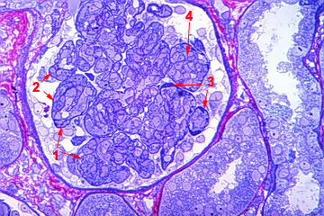
Fig 1. MPGN I with MPC, mesangial cell proliferation and
subendothelial immunoglobulins.
GNMP I con CMP, proliferación mesangial y depósitos
inmunes sub endoteliales.
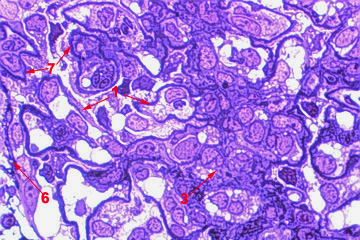
Fig 2.Dense Deposit Disease without MPC but with mesangial cell proliferation.
Glomerulopatía por depósitos densos sin CMP pero con proliferación messangial.
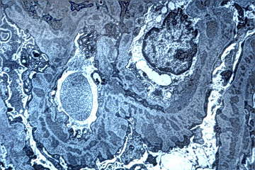
Fig 3. Membranous GN with deep penetration of deposits and MPC.
GN membranosa con penetración profunda de los depósitos epimembranosos y CMP.
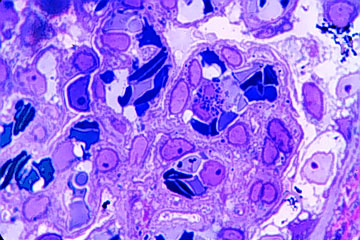
Fig 4. MPC in sickle cell disease.
CMP en enfermedad de células falciformes
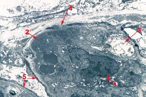
Fig 5. FSGS with segmental hyalinosis and segmental MPC.
EGFS con hyalinosis segmentaria y CMP.
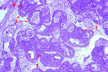
Fig 6. MPC with no identifyable subendothelial deposits. Etiology unknown.
CMP sin depósitos subendoteliales identificables. Etiología desconocida.
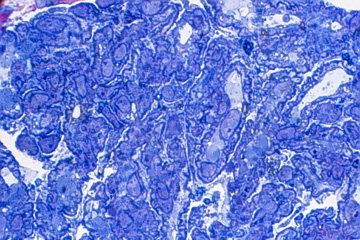
Fig 7. Thrombotoic micoangiopathy, MPC and subendothelial fibrinogen.
Microangiopatía trombótica, CMP y fibrinógeno subendotelial.
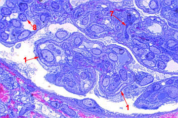
Fig 8.Chronic rejection glomerulopathy with MPC, no deposits.
Glomerulopatía del trasplante crónico con CMP, no depósitos.
REFERENCES / REFERENCIAS: