
PREVALENCE OF LEFT VENTRICULAR HYPERTROPHY AND GEOMETRIC MODELLING IN PATIENTS WITH CHRONIC RENAL FAILURE
N.A.Tomilina, G.V.Volgina, B.T.Bikbov, Yu.V.Perepechyonickh, I.I.Stenina
E-mail: Volgina Galina Vladimirovna
Moscow. Russia
| DISCUSSION BOARD |
Abstract
Background. Left ventricular hypertrophy (LVH) is an independent risk factor for morbidity and mortality in patients with CRF. Prevalence, risk factors of LVH and geometric models of left ventricle were evaluated in 150 patients with CRF and in 160 patients with ESRD.
Methods. LVH was defined as an abnormal increase in left ventricular mass (LVMI) is detected by echocardiography in patients with CRF.
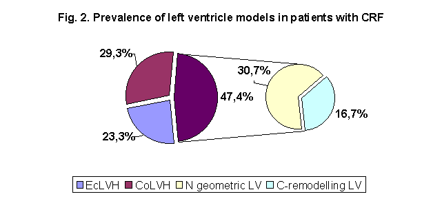
Results. It was observed in 52.6% of predialysis patients. In the group of patients with Ccr 50-70 ml/min it was diagnosed in 26.4% of cases, in those with Ccr 49-25 ml/min - in 46.3% of cases, and in patients with Ccr < 24 ml/min - in 68% of cases. Concentric LVH was observed in 29.3% in the predialysis group with LVH, eccentric LVH was in 23.3%. In the group of HD patients LVH was detected in 78.1%: concentric LVH was found in 51.8% and eccentric LVH was present in 26.3%. In patients without LVH we found concentric remodeling of left ventricle in 16.7% of predialysis patients and in 13.1% of HD patients (p<0.05). We found that risk factors for LVH were the decline in Ccr (p<0.05), age (p<0.001), increase of blood pressure (p<0.0001), anemia (p<0.04). In HD patients additional risk factors were: levels phosphorus (p<0.0001), product CaxP (p<0.0001) and interdialytic weight gain (p<0.05). Risk factors for concentric remodeling were age (p<0.05), duration of predialysis hypertension (p<0.04), creatinine plasma level (p<0.05), anemia (p<0.005).
Conclusions.LVH is a frequant finding in patients with CRF. Prevalence of LVH increases with decline of renal function. Risk factors for LHV at this stage of kidney disease are age, high blood pressure, anemia. The same mechanisms are accounted for LVH in dialysis patients. Risk factors for LHV in patients with ESRD are high blood pressure, anemia, levels phosphorus and product CaxP. Fluid overload may be considered an additional important risk factor of LVH in HD-patients. The prevalence of LVH in CAPD-patients is less than in HD-patients. The dominant of LV geometric models in the observed cohort of dialysis patients was CoLVH. C-remodelling LV can develope in patients with normal myocardial mass. We suggest that C-remodelling LV represents the early stage of the development of LVH.
Keywords: chronic renal failure, end-stage renal disease, left ventricular hypertrophy, geometric modelling left ventricle
INTRODUCTION
Left ventricular hypertrophy (LVH) represents the main manifistation of uremic cardiomyopathy. It is considered a predictor of both cardiovascular events and death in patients with ESRD, regardless of age, diabetes mellitus, hypertension, hyperlypidemia, smoking and coronary heart disease [1-5].
Myocardial mass gain is not the only factor, which has an impact on clinical outcome. The different geometric models (concentric remodelling, concentric or eccentric LVH) play an important role as well [6-11,18]. Concentric LVH (CoLVH) depends primary on persistence pressure overload and artheriosclerosis. These factors launch process of myocardial cells involvement and fibroblasts proliferation, while the left ventricle volume increases nonsignificantly. The volume overload caused by sodium and water retention anemia and arteriovenous fistula results in eccentric LVH (EcLVH), i.e. dilatation of left ventricle (LV) volume accompanied by myocites elongation [11-16].
Risk of cardiovascular events is the highest in patients with CoLVH. It is smaller in cases of EcLVH and it is minimal in patients with concentric remodeling (C- remodeling) [7,9,10,17].
The data of the prevalence of LVH in chronic renal failure (CRF) are controversial. It was shown in 25% to 87% of predialysis patients and in 50% to 97% of dialysis patients [18-26]. There is no agreement about the frequency of LVH geometric models in ESRD. While the most studies demonstrate predomination of CoLVH (40-63% versus 20-30% of EcLVH) [14,22,26], some authors report the higher prevalence (63-79.6%) EcLVH [20,23,27].
The aim of the study is to estimate the prevalence of LVH and its geometric models in patients with CRF and to investigate the risk factors of myocardial remodeling in ESRD.
Subjects and methods
Three hundred ten patients (150 patients with CRF and 160 dialysis patients) were enrolled in the study (Table 1). Ecxlusion criteria were multisystem disease, diabetes mellitus, severe cardiac valvular disease, congestive cardiac failure, amyloidosis, inadequate echocardiographic images.
In the group of dialysis patients 130 (66 male, 64 female) were treated with hemodialysis (HD) and 30 (15 male, 15 female) were CAPD-patients. The average age of HD- and CAPD-patients was almost the same: 49,35±13,6 years (range 18-73) and 51,8±16,0 years (range 20-76) respectively. Duration of dialysis treatment was 29,4±27,3 months (range 1 - 121, mediana 19,0) in the subgroup of HD-patients and 15,5±16,2 month (range 1 - 45, mediana 6,5) in the subgroup of CAPD-patients.
Table 1. Demographics data and causes of chronic renal failure in predialysis and dialysis patients
| Predialysis patients (n=150) | Dialysis patients (n=160) | |
| Age, years | 56,2±13.1 (26-79) | 49.6 ±15.4 (18-76) |
| Males/females, n (%) | 78/72 (52/48) | 81/79 (50,6/49,4) |
| Causes of CRF, n (%) | ||
| Glomerulonephritis | 74 (49,3) | 74 (46,2) |
| Hypertensive nephrosclerosis | 8 (5,3) | 14 (8,7) |
| Polycystic kidney disease | 15 (10) | 20 (12,5) |
| Obstructive uropathy | 15 (10) | 10 (6,3) |
| Tubulointersticial nephritis | 3 (2,0) | 3 (1,9) |
| Pyelonephritis | 23 (15,3) | 16 (10,0) |
| Other nephropathy | 7 (5,0) | 15 (9,4) |
| Unknow nephropathy | 5 (3,3) | 8 (5,0) |
The role of age, anemia, hypertension and its duration, as well as degree of CRF (estimated by creatinine clearance), serum albumin level, hyperphosphataemia, secondary hyperparathyreoidism and dialysis modality was investigated. In 130 HD-patients the average interdialytic weight gain has been analyzed as well.
Hemoglobin (Hb), serum electrolytes, serum creatinine (Cr), albumin, calcium, phosphate, calcium-phosphate product, as well as alkaline phosphatase and intact parathyroid hormone (iPTH) were evalueted. The Cоckcroft - Gault formula was used to calculate creatinine clearance (Ccr) [28].
Echocardiography was performed using "Aloca SSD-2000"(Japan) with a 3-MHz mechanical sector transducter. Measurements were made according to the recommendations of the American Society of Echocardiography [29]. Echocardiography was performed within 18 to 24 hours after routine dialysis in HD-patients, and after the dialysate removal in CAPD-patients. The following echocardiographic data were evalueted: left ventricular end diastolic dimention (LVEDD), left ventricular end systolic dimention (LVESD), left ventricular posterior wall thickness (PWT), interventricular septal thickness (IVS), left atrial dimention (LA). Relative wall thickness (RWT) was calculated: RWT = IVS+PWT/LVIDD. Left ventricular mass was determined by M-mode echocardiography using the following formula: LVM (g) = 0.8*1.04{(LVIDD + PWT + IVS)3 – (LVIDD)3} + 0,6 [30,31]. LVM was indexed per square metre of body surface area (BSA). Left ventricular size was measured at just below the trips of the mitral valve leafleta at the largest left ventricular internal dimention. LVEDD and PWT measurement were made at end-diastole. Left ventricular hypertrophy (LVH) was defined as 2 SD above the mean LVMI found in the Framingham Study population. Left ventricular hypertrophy was defined in absolute terms as more than 134 g/m2 in men and more than 110 g/m2 in women. Concentric hypertrophy was diagnosed when LVH was accompanied by RWT greater than 0,45. Eccentric hypertrophy was defined in cases of LVH and RWT less than 0,45. C- remodelling was defined when the myocardial mass was normal but RWT was more than 0,45. Hypertension was diagnosed when the mean BP (MBP) was higher than 105 mm Hg or systolic/diastolic BP (SBP/DBP) was higher than 140 mm Hg/ 90 mm Hg, respectively [32-34].
Statistical methods
For normally distributed continuous variables, mean values and standart deviations were calculated. For variables with skewed distribution, median values and interquartile ranges were determined. Statistical comparison of continuous variables was carried out sing Kruskal-Wallis and Mann Whitney U-test. Comparison of qualitive variables and proportions were performed using chi-square and for analysis of 2*2 contingency tables with two-tailed Fisher’s exact test. Correlations were sought using the Spearman correlation test. A P value of < 0,05 was considered to be statisticaly significant. Statistical analysis was performed using the computer software SPSS 8.0 (SPSS Inc., Chicago).
Results
Prevalence of LVH and its geometric models in patiets with CRF
The average prevalence of LVH in the whole group of patients with CRF was 52,6% (Figure1). Figure 1 shows that there was the close correlation between Ccr and the frequency of LVH.
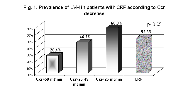
On the other hand the correlation between the prevalence of LVH and both Hb level and age was found (Table 2). But it is noteworthy, that LVH was observed in 26,4% of patients with Ccr above 50 ml/min in spite of normal average Hb level in this subgroup. There was no correlation between the prevalence of LVH and BP, but it should be noticed that all the patients were hypertensive. Clinical and echocardiograpic parameters in patients without LVH (LVH-) and with LVH (LVH+) are shown in the Table 3. Predialysis patients with LVH were older, had higher serum creatinine level, MAP, systolic (but not diastolic) BP. Patients without LVH had higher Нв and Нt levels.
Table 2. Clinical, laboratory and echocardiographic parameters in patients with chronic renal failure
| Parametrs | CRF | P value | |||
| In total (n=150) | Ccr 75 - 50 ml/min (n=34) | Ccr 49 - 25 ml/min (n=41) | Ccr <25 ml/min (n=75) | ||
| Age, years | 52.2±13.1 | 41.5±7.1 | 48.0±10.1 | 57.8±14.2 | 0.001 |
| 94/56 | 19/15 | 26/15 | 49/26 | 0.91 | |
| BMI, kg/m² | 26.0±5.3 | 27.6±8.6 | 25.8±3.8 | 25.7±2.4 | 0.9 |
| Hemoglobin, g/l | 105.0±25.3 | 136.3±16.5 | 110.2±22.2 | 82.5±23.8 | 0.001 |
| SBP, mm Hg | 160.3±21.9 | 148,5±15.2 | 146.7±17.75 | 160.2±23.5 | 0.4 |
| DBP, mm Hg | 89.8±12.7 | 91.1±13.6 | 90.8±9.25 | 85.0±10.8 | 0.36 |
| MBP, mm Hg | 110.9±14.3 | 107.2±15,0 | 109.4±10.1 | 109.9±13.5 | 0.8 |
| LVM, g | 292.8±102.8 | 203.5±94.0 | 292.0±76.5 | 322.6±118.6 | 0.0001 |
| LVMI, g/m² | 159.5±49.2 | 103.35±49.6 | 153.1±36.75 | 184.1±57.0 | 0.0001 |
| LVH, % | 52.7 | 26.4 | 46.3 | 68.0 | 0.001 |
Note. CRF – chronic renal failure; BMI – body mass index; SBP – systolic blood pressure; DBP – diastolic blood pressure; MBP – mean blood pressure; LVM – left ventricular mass; LVMI - left ventricular mass index; LVH – left ventricular hypertrophy
Table 3. Clinical and echocardiographic parameters in patients with chronic renal failure without LVH (LVH-) and with LVH (LVH+)
| Parametrs | CRF | P value | |
| (LVH -) | (LVH+) | ||
| LVMI, g/m² | 96,0±16,2 | 174,4±41,85 | 0,0001 |
| Age, years | 47,4±4,3 | 58,0±12,6 | 0,005 |
| Cr, mmol/l | 272,0 (147,5; 376,2) | 472,0 (180,0; 810,0) | 0,01 |
| SBP, mm Hg | 141,25±13,5 | 153,5±24,7 | 0,02 |
| DBP, mm Hg | 88,75±6,4 | 92,35±13,6 | 0,15 |
| MBP, mm Hg | 106,25±8,0 | 112,75±15,8 | 0,036 |
| Duration of hypertension, months | 0,67 | ||
| BMI, kg/m² | 26,8±6,2 | 25,8±4,45 | 0,62 |
| Hb, g/L | 118,2±18,4 | 97,8±26,45 | 0,05 |
| Ht, % | 36,55±2,6 | 32,4±2,4 | 0,05 |
| Albumin, g/L | 41,4±3,4 | 38,7±5,0 | 0,24 |
| Ca, mmol/L | 2,1±0,35 | 2,25±0,20 | 0,21 |
| P, mmol/L | 1,75±0,41 | 1,74±0,59 | 0,88 |
| CaxP, mmol/L | 4,89±1,08 | 4,75±1,02 | 0,87 |
| iPTH, pg/ml | 293,1±256,4 | 274,3±242,8 | 0,99 |
According to our data patients with LVH were older (p<0,005), had higher levels of serum creatinine (p<0,01), SBP (p<0,02) and MBP (p<0,036), but not DBP.
Figure 2 shows the prevalence of LVH geometric models in patients with CRF. CoLVH was observed in 23,3% of them and EcLVH was diagnosed in 29,3% patients. LVMI was in normal range in 47,4% patients with CRF, but C- remodeling was found about in one third of them (in 16,7% of patients).
Conclusion
The prevalence of LVH in CRF is close related with the degree of Ccr decrease. The risk factors of LVH in CRF are hypertension, anemia and age, but we suggest that each of these factors may play different role on different stages of CRF. While hypertension may be considered one of the initial risk factors of LVH, anemia may play the most important role at the time of CRF progression. The prevalence of CoLVH and EcLVH in CRF is equal, and C-remodelling of LV can be defined in one third of patients with normal LVMI.
Figure 3 shows the prevalence of LVH in the whole group of dialysis patients as well as its frequency according to dialysis modality. It was found that the prevalence of LVH was significantly higher in HD-patients in comparison to CAPD-patients (79.4% and 63.6% respectively, p<0.05).
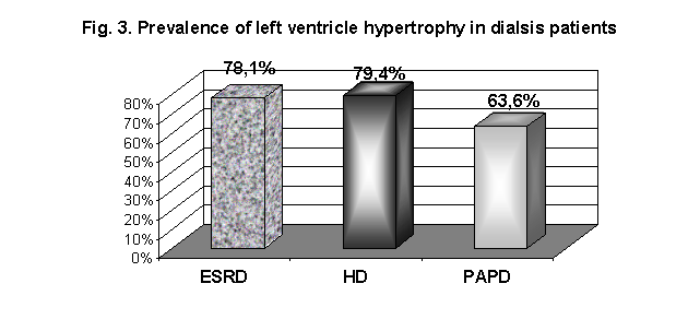
In order to study risk factors of LVH in dialysis patients we have analysed clinical and biochemical parameters in two subgroups of these patients divided according to LVMI (Table 4). Dialysis patients with LVH were significantly older and had higher levels of BP. It should be noticed that although we could not found difference between average Hb level in both subgroups of patients anemia observed more frequent in patients with LVH (p< 0,05).
Table 4. Clinical and biochemical parameters in subgroups of dialysis patients divided according to LVMI.
| Parameters | ESRD | ||
| (LVH -) | (LVH+) | P value | |
| LVMI, g/m² | 102.9 ± 17.7 | 172.8 ± 42.4 | 0.0001 |
| Age, years | 44.9±13.9 | 50.7±13.6 | 0.048 |
| Duration of dialysis treatment, months | 23.4±22.8 | 30.8±32.4 | 0,35 |
| Interdialytic weight gain, kg | 1.86±1.15 | 2.43±1.0 | 0.047 |
| SBP, mm Hg | 127.9±17.35 | 149.0±18.2 | 0,002 |
| DBP, mm Hg | 78.6±9.5 | 89.4±10.25 | 0,009 |
| MAP, mm Hg | 95.1±11.6 | 105.6±11.8 | 0,003 |
| Duration of hypertension, months | 158.7±120.5 | 170.0 ± 135.3 | 0,67 |
| Hb, g/L | 97.7±22.8 | 91.7±20.8 | 0,11 |
| Albumin, g/L | 36.5±3.55 | 35.75±5.2 | 0,56 |
| Ca, mmol/L | 2.38±0.15 | 2.36±0.22 | 0,87 |
| P, mmol/L | 2.0±0.4 | 2,07±0.45 | 0,23 |
| CaxP, mmol/L | 4.81±1.17 | 4.81±1.05 | 0,42 |
| iPTH, pg/ml | 0,11 | ||
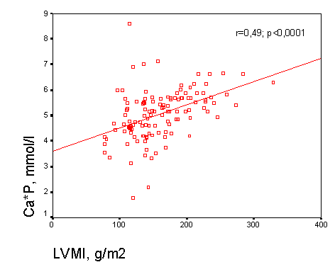
The positive correlation between LVMI and SBP (Figure 4) (r=0,45; p<0,0001), DBP (Figure 5) (r=0,44; p<0,0001), phosphorus (Figure 6) (r=0,45; p<0,0001), product CaxP (Figure7) (r=0,49; p<0,0001), as well as the negative correlation between LVH and Hb (r=-0,38; p<0,04) were found.
Besides LVH was defined more often in patients who were treated with acetate HD versus those on bicarbonate HD (χ²=3,95, р
<0,047).Fig. 4. Correlation between LVMI and SBP
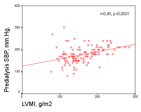
Fig. 5. Correlation between LVMI and DBP
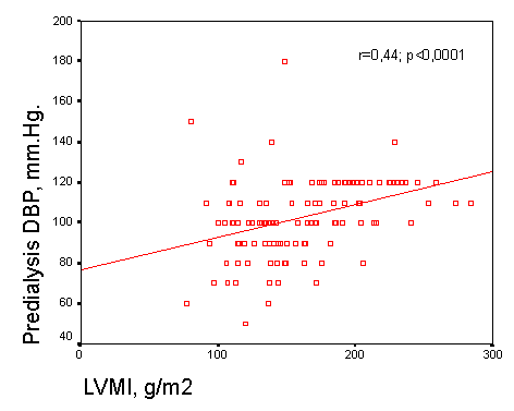
Fig. 6. Correlation between LVMI and phosphorus
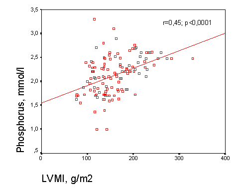
Fig.7. Correlation between LVMI and product CaxP
The prevalence of different geometric models in the group of dialysis patients is shown on the Figure 8.
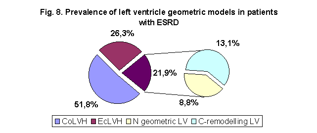
CoLVH was diagnosed in 51,8% of them and EcLVH was found only in 26,3% of dialysis patients. In 21,9% of patients LVMI was not increased, but C-remodelling was revealed almost in two thirds of them. So the proportion of Ec LVH in dialysis patients in compare to predialysis group was decreased and we suggest that the greater volume overload may be accounted for this decline. The prevalence of C-remodelling in dialysis group was higher than in predialysis patients
Conclusion
The prevalence of LVH in dialysis patients is the highest on HD-treatment. The most important risk factors in ESRD are age, hypertension, anemia, levels phosphorus, product CaxP, as well as fluid overload and fluid overload with increased interdialytic weight gain (increase of extracellular blood volume) are risk factors of LVH in patients with ESRD. In observed cohort of dialysis patients we have found predomination of CoLVH. The proportion of patients with C-remodelling among thouse of tem who had normal LVMI was significantly less then in the subgroup of predialysis patients.
In order to study the risk factors of C-remodelling of left ventricule we have analysed clinical data of two subgroups of patients with normal range of LVMI divided according to geometry of LV (Table 5). It was found that patients with C-remodelling in comparison to thouse who had normal LV geometry were significantly, more hypertensive with longer hipertension history. In dilysis patients with LV C-remodelling greater average interdialytic weight gain was revealed as well (Table 5).
Table 5. Demographic and clinical parameters in subgroups of patients with normal with normal left ventricular geometry and C- remodeling of left ventricule
| Parametrs | |||
| LVMI, g/m² | 90.0±20.75 | 103.5±15.7 | 0.01 |
| Age, years | 36.0 ± 9.0 | 50.6±14.2 | 0.005 |
| Males, % | 52 | 57 | NS |
| Duration of dialysis treatment, months | 19.0(2.5; 33.0) | 18.0 (1.0; 44.0) | 0,86 |
| Interdialytic weight gain, kg | 1.33±1.35 | 2.33±0.78 | 0.05 |
| Duration of hypertension, months | 89.9 ± 86.55 | 190.8 ± 122.8 | 0.04 |
| SBP, mm Hg | 137.5±16.6 | 148.2±18.4 | 0.01 |
| DBP, mm Hg | 84.2±10.0 | 93.2±9.5 | 0.01 |
| Hb, g/L | 96.9±21.3 | 88.2±24.4 | 0.05 |
Summary
Our study demonstrates that LVH is a frequant finding in patients with CRF which proportion increases with aggravation of renal dysfunction. These data let us confirm that mechanisms of cardiovascular disease persist long before starting dialysis. Risk factors for LHV at this stage of kidney disease are age, high blood pressure, anemia. Prevalence of LVH increases with decline of renal function. The same mechanisms are accounted for LVH in dialysis patients. Risk factors for LHV in patients with ESRD are high blood pressure, anemia, levels phosphorus and product CaxP. Fluid overload may be considered an additional important risk factor of LVH in HD-patients. The prevalence of LVH in CAPD-patients is less than in HD-patients. This difference may be explained by hemodinamic conditions and better compliance of blood pressure on this type of dialysis.
Prevalence of CoLVH and EcLVH in patients with CRF was quite simillar (29.3% and 23.3%). The dominant of LV geometric models in the observed cohort of dialysis patients was CoLVH. C-remodelling LV can develope even in patients with normal myocardial mass. It was found in CRF and its prevalence increased with decline of renal function. We suggest that C-remodelling LV represents the early stage of the development of LVH.
REFERENCES