|
|
The usefulness of the intraoperative Power Doppler ultrasound in Neurosurgery. (preliminary report)
Authors: Latka Dariusz, Kita Pawel, Czerwinski Krzysztof, Hendryk Stanislaw*, Mrowka Ryszard* *Chair and Department of Neurosurgery, Silesian Medical Academy, Bytom, Poland Institution: Department of Neurosurgery, State Medical Center, Opole, Poland Email: dlatka@wcm.opole.pl Abstract Introduction Materials & Methods Results & Discussion Modern neurosurgery requires intraoperative image techniques. The aim of study is to estimate efficacy of Power Doppler in the intraoperative evaluation of brain lesions. In the examined period the intraoperative ultrasound was performed in over 10% of all patients operated on in our Department. Color and Power Doppler B-mode sonography offers new possibilities of intraoperative control of neurosurgical procedures - we use it mostly in the recognition of tumors’ vascularization, small AVMs’ review and in the estimation of proper vascular clip application in aneurysm surgery. The ultrasonographic technique has been used in 54 tumors from among 126, in 5 from 6 abscesses, in 6 from 40 intracerebral hematomas, in 2 from 72 aneurysms and in 1 from 2 AVMs procedures. The ultrasound imaging using Power Doppler technique appears to be of high quality for therapeutic decision making in the procedures requiring vascular estimations. Its enables the fantastic estimation of tumors vascularization, helps to avoid unnecessary bleeding from earlier detected supplying vessels and to preserve vessels being in contact with tumor. No intraoperative complications and no postoperative infections result from use of ultrasound in the operating room. Our procedures are as fast as before, safer by virtue of informations obtained by ultrasonographic intraoperative examination. The Power Doppler modality enables the fantastic estimation of tumors vascularization, helps to avoid unnecessary bleeding from earlier detected supplying vessels and to preserve vessels being in contact with tumor. It also seems to be very useful in proper clips placement in vascular surgery. A very expensive techniques of intraoperative image guidance like open-angioMR, intraoperative angiography or computer assisted neuronavigation are being used to give the neurosurgeon better knowledge about the brain vascularisation in the operation theatre. Intraoperative ultrasound using Power Doppler modality is a technique which is becoming to be accepted since early nineties with the development of modern ultrasonographic devices. It is much less expensive technique and according to many authors it gives the images of a comparable to conventional angiography images. The aim of this study is to preliminary present our own experience in using of this modality and to discuss the future perspectives for Power Doppler in neurosurgical intraoperative diagnostics armamentarium. Till January'99 the intraoperative ultrasound imagining was performed in 68 patients operated on in the Department of Neurosurgery in Opole – it means that this technique was employed in over 10% of all neurosurgical procedures (total number of operations during this period was 650). We have been using the Panther 2002 - it is a very sophisticated machine working in the simple real-time B-mode as well as giving the possibility of duplex and triplex presentations using Color and Power Doppler modes. It is equipped with the multifrequency 5,5-7,5 MHz probes designed especially for open neurosurgical applications. The access to the intraoperative ultrasound is absolutely full in our Department - it depends only on the operator will; he takes it when he supposes it to be useful. It appeared that this technique has been used in 54 tumors from among 126, in 5 from 6 abscesses, in 6 from 40 intracerebral hematomas, in 2 from 72 aneurysms and in 1 from 2 AVMs. 54 patients with brain tumors intraoperatively ultrasonographically scanned were included to the comparative study with the group of 70 brain tumor cases operated on without the use of ultrasonography. Such features as: the type of tumor, localization (either its’ depth or the brain area involved) and tumor vascularization or the nearness of important vascular structures have been taken into consideration. These data have been analyzed statistically using standard correlative procedures (chi-squared analysis) in hope to find out answer which of the indications to use this technique are positively verified by a everyday practice. B-mode real-time linear sonographic scanning can be used to localize central nervous system lesions such as hematomas, tumors, cysts, abscesses or vascular malformations. Depending on our experience the resolution obtained with ultrasonography seems to be similar to that of CT and the echogenic pattern of the normal brain parenchyma can be easily differentiated from the abnormal pattern of the lesion. It gives very few obstructing artifacts. Most of the lesions give at least partly higher echosignals than normal brain tissue, except for cysts and abscesses (usually hypoechogenic). In all of our cases the investigated lesions could be identified during intraoperative ultrasound investigations and in almost all of them the patomorphological features like size and shape were comparable with preoperative CT findings. In some cases we found it even more accurate than CT in the estimation of unclear boundary between the tumor and the brain edema: in benign gliomas the CT sometimes gives a misleading picture of more or less clearly delineated tumor. Ultrasonography also allowed more accurate differentiation between intratumoral necrosis and cysts than CT. In our series intraoperative ultrasound performed on the dura-mater or on the cortex has proven to be extremely useful for detecting and localizing subcortical rather than deep-seated brain tumors as well as hyperechogenic intraparenchymal hematomas. The deep-seated tumors were also detected with accuracy, but this fact allows rather ultrasonographically guided biopsy than open approach to the lesion. We almost haven’t used ultrasonography in skull base tumors; neither meningiomas (except the ones situated close to important vessels or sinuses), nor neurinomas, hypophysis adenomas or craniopharyngeomas. Very rarely we used this device in posterior fossa lesions. Maximum usage of ultrasound takes place during gliomas (both: benign and malignant) and metastases operations because of their subcortical localization. The advantages of the technique were particularly evident when searching for tumors which were not visible from the brain surface. In these conditions intraoperative sonography greatly facilitates the planning of the operative approach. Choosing this optimal approach to the tumor, the surrounding brain is minimally damaged. The usefulness of the application of Power Doppler mode in tumor vascularization estimations must be outlined. It helps to avoid unnecessary bleeding from earlier detected supplying vessels and to preserve vessels being in contact with the tumor (Figure 1).
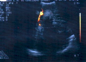
Figure 1 A case of tentorial meningioma adherent to the sagittal sinus. In the Power Doppler presentation you can see the tumor in the relation to the sinus.
Ultrasound B-mode Doppler sonography with high resolution probes offers new possibilities of intraoperative control of neurovascular procedures - our experience in this subject is modest - we suppose it can be useful in the localization of small arteriovenous malformations (AVMs) and feeders estimation.
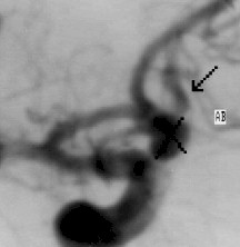
Figure2 A case of parasagittal parietal AVM. The Power Doppler sonography gives the image comparable to angiography.
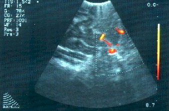
During the treatment of aneurysm prior to clipping you can find whether blood flows through (Fig. 3), and after clipping it is possible to evaluate whether artery is still permeable without stenosis or torsion.
Figure 3 A case of Aco-A saccular aneurysm in the Power Doppler sonography prior to the clip application.
No intraoperative complications and no postoperative infections result from the use of ultrasound in the operating room. Surgical procedures, as a result, are as fast as before, safer and better planned by virtue of information obtained by ultrasonographic intraoperative examination, instead of simple preoperative imaging procedures. Thanks to the relatively low-cost, non-invasive character, and simplicity of use, the intraoperative Power Doppler ultrasound should be highly recommended in a number of practical applications in neurosurgical practice and will be a subject for our further estimations.
References in authors possession. |
To contact
Authors personally: dlatka@wcm.opole.pl |
|