|
|
POSTTRAUMATIC (?) HEMANGIOENDOTHELIOMA OF THE CALVARIAL REGION. PRELIMINARY CASE REPORT
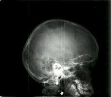
Authors: Kita Pawel, Latka Dariusz, Czerwinski Krzysztof, Szczurek Zbigniew* * Chair and Departament of Pathology Silesian Medical Academy Zabrze, Poland Institution: Department of Neurosurgery, State Medical Center, Opole, Poland Email: pkita@wcm.opole.pl Abstract Introduction Case Report Discussion WE REPORT THE: 11-year old girl who underwent excision of an intracranial extradural tumor of the right calvarial area. The evidence of that case is connected with the head trauma which occurred two months before the appearance of the right calvarial area tumor. Total excision of the tumor was made and histologically diagnosed as epithelioid hemangioendothelioma. INTRODUCTION Hemangioendotheliomas are heterogenous group of rare tumors. With respect to their histological appearance and their biological behaviour these tumors are intermediate between benign hemangiomas and malignant angiosarcomas. There are recognised four subgroups of these tumors: the epithelioid hemangioendothelioma, a spindle cell variant, kaposiform hemangioendothelioma and malignant endovascular papillary angioendothelioma, also known as a Dabska tumor. CASE REPORT An 11-year old girl was admitted to the hospital for the treatment of tumor on the calvarial area over the right parietal region. The tumor caused the destruction of the skull and penetrated its interior. It didn’t interfere in the brain. In the family history the parents associate the growth of this outgrowth with the head injury in the summer this year. After the head injury there appeared a hard thickening painful at palpation. It was removed by surgeon from under the skin surface. It was recognised as a hematoma. After about a month the reappearance of the tumor was observed on the calvary and this time it was much bigger. Because of its dynamic growth conventional radiogram of skull (Figure 1) and CT (Figure 2) were made.
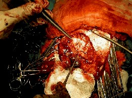
At admission to our hospital the girl was in a good state without any neurological signs. Locally a tumorous change 4x5cm large on the right parietal region and hard skin non movable over the lesion. The girl was operated on and the lesion was totally removed. This lesion didn’t infiltrate dura or the skin (Fig.3). In the place of the removed bone a Cranioplast plate was fixed (Fig.4). The postsurgical recovery was uncomplicated. The girl was qualified to post-operative radiotherapy. Released home in good state.
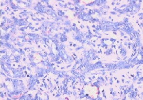
HISTOLOGICAL FINDINGS
Among the hyaline eosynophylic stroma occur multishaped chaotically situated vascular channels lined with irregular endothelial cells. Occasionally to be found bigger vessels of mature structure resembling small veins. These changes are accompanied in the stroma by mostly disseminated or bigger concentrations of lymphocytic cells (Fig5). Under greater magnification inside some vascular channels solitary erythrocytes were found. Endothelial cells had longitudinal spindle- shaped nuclei. In some cells a bright extension of vacuolar nature was observed indicating the epithelial nature of the cells (Fig6). In other areas of the tumor in the stroma more or less numerous erythrocytes were observed. No bone tissue, focal necrosis, myxoidal changes or cartilaginous differentiation were in the tumor. No mitotic activity was observed. DISCUSION The epithelioid subtype is the most commonly described entity within the central nervous system, meningeal coverings or skull (9) First described by Weiss and Enzinger (7). Most cases of hemangioendotheliomas are reported in liver (4,5), lungs (2), digestive system (7), head and neck (3,7), bone(1,6) and heart (4) Epithelioid hemangioendothelioma is rarely observed in children (9) The tumor occurs in superficial or deep soft tissue and is described as a single mass, slightly painful at palpation. In many cases the tumor is related to vessels especially to veins (7,8). According to Enzinger: "The tumor are composed of short strands or solid nests of rounded to slightly spindled endothelial cells. Rarely are large, distinct vascular channels seen, except in the more peripheral portions of the tumor.Instead the tumor cells form small intracellular lumina, which are seen as clear spaces or „vacuoles" that distort or „blister" the cell. [...] The stroma varies from highly myxoid to hyaline. [...] in most cases the tumors appear quite bland and there is virtually no mitotic activity. In about one fourth cases the tumors contain areas with significant atypia, mitotic activity (greater than 1/10HPF), focal spindling of the cells, or necrosis. Such features can be correlated with a more aggressive course."(8) The differential diagnosis includes metastatic carcinoma or melanoma, and also many sarcomas that may adopt an epithelioid appearance. In uncertain cases immunohistochemistry as well as electron microscopy may be helpful in the differentiation.(8). The overall prognosis of epithelioid hemangioendothelioma, although no yet well defined, seems quite favourable. In the period of 4 years 13% of the patients noted recurrence, 31% developed metastasis, and the mortality rate was 13% (8). Treatment of this tumor should comprise wide surgical excision. The role of adjuvant radiotherapy or chemotherapy is not well established now. REFERENCES
|
To contact
Authors personally: pkita@wcm.opole.pl |
|