| Paper # 001 | Versión en Español  |
Marcial Garcia-Rojo, Jesús González, Ana Morillo, Jesús Martín
[Title] [Introduction] [Results] [Pictures] [Discussion] [Bibliography]
We describe a 72 years old man that came into hospital
suffering from asthenia and a 7 Kilograms weight loss. In his
personal history, he suffered  from spondylo-osteoarthritis and a
prostatectomy for a benign prostate hyperplasia. At that time he
has a good general status, without fever and with a normal
pulmonary and cardiac examination. He was discovered to have
multiple adenopathies in neck, axilla and inguinal regions, the
biggest one was of 3 cm. In laboratory tests, no significant
alterations were found. Hemoglobin as 13 g/dl and leukocytes
count was 10.500 per microliter with 29.9 % segmented and 60.7%
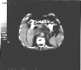
of lymphocytes. CT-Scan showed multiple adenopathies in
mediastinum, retroperitoneum, mesenterium and around iliac and
femoral vessels bilaterally.
A biopsy was performed from an adenopathy of the axillary region.
The histopathological examination showed it was a low grade
B-cell non Hodgkin lymphoma type lymphoplasmacytoid
(immunocytoma).
A few days later of the diagnosis of lymphoma he was started on
CVP (Cyclophosphamide 600 mg per day, Vincristine 1.5 mg per
square meter in bolus and Prednisone 90 mg per day) for five
days. Allopurinol and gastric protection were also prescribed.
After five cycles of chemotherapy the response to these agents
was poor. Peripheral blood study after the last cycle was:
leukocytes 8100 per microliter with 29.1% segmented and 53.3 % of
lymphocytes. Hemoglobin was 13.6 g/dl.
 Six
months after the diagnosis of lymphoma the patient began with
epigastric pain, pyrosis and vomiting of increasing intensity. A
gastroduodenal radiological study was performed and it showed an
stenosis and lack of distention at fundic area, with an irregular
pattern of the mucosa, suggesting an malignant process. Gastric
endoscopy revealed an normal esophagus, with a rigid gastric
fundic area showing enlarged and irregular foldings of the mucosa
that reached the antrum, bleeding very easily. Pylorus and
Duodenum were normal. Several biopsies were taken from fundic
area and antrum.
The histopathological studies of the gastric biopsies revealed
the presence of a gastric signet ring adenocarcinoma.
At that time a new CT scan study showed the persistence of all
the adenopathies found in the first study. No hepatic or
pulmonary metastases were found.
A total gastrectomy and splenectomy were performed. At the time
of surgery, multiple adenopathies were noted in the proximity of
the head of the pancreas and in the lesser omentum and
gastrocolic omentum. No hepatic metastases were noted. No
postoperative complication presented.
Pathological procedures
Lymph node specimens were fixed in B5 solution. The rest of the
specimens were fixed in buffered 10% formalin. Paraffin-embedded
tissue sections were stained immunohistochemically by the
streptavidin-biotine-alkaline phosphatase method with antibodies
Common Leukocyte Antigen (CLA, Biomeda), CD-20 (L-26, Biomeda),
CD45R0 (UCHL-1, Biogenex), Kappa light chain (Kappa, Biomeda),
Lambda light chain (Lambda, Biomeda), keratin (CK8, 18 & 19,
Biogenex) and Common Epithelial Antigen (CEA, Biomeda). The
staining method for keratin required pretreatment with enzymes
and microwave heating.