| Paper # 095 | Versión
en Español |
| Paper # 095 | Versión
en Español |
Andréa Rodrigues Cordovil Pires, Luciana Wernersbach Pinto.
[Title] [Introduction] [Materials and Methods] [Results] [Discussion] [Bibliography]
|
|
|
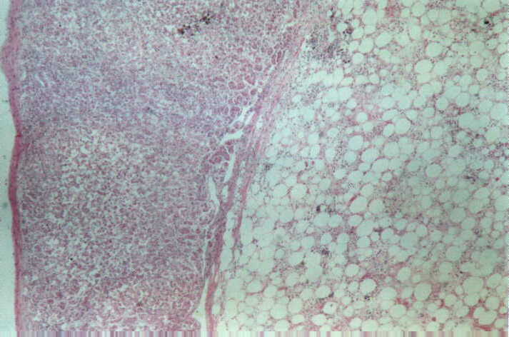
Left adrenal myelolipoma. The outer layer on left side is represented by compressed adrenal cortical cells.In the right side there are fat cells and trilineage hematopoietic cells. HE, 40x. |
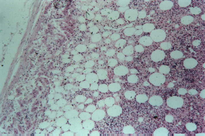 |
Left adrenal myelolipoma. The outer layer on left side is represented by compressed adrenal cortical cells.In the right side there are fat cells and trilineage hematopoietic cells. HE, 100x. |
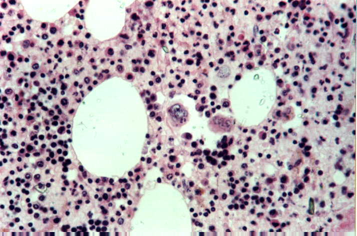 |
Left adrenal myelolipoma. Close view of fat cells and trilineage hematopoietic cells. There are megakaryocyte, erithrocyte and myeloid lineages in all stages of differentiation, all of them typical. HE, 40x. |
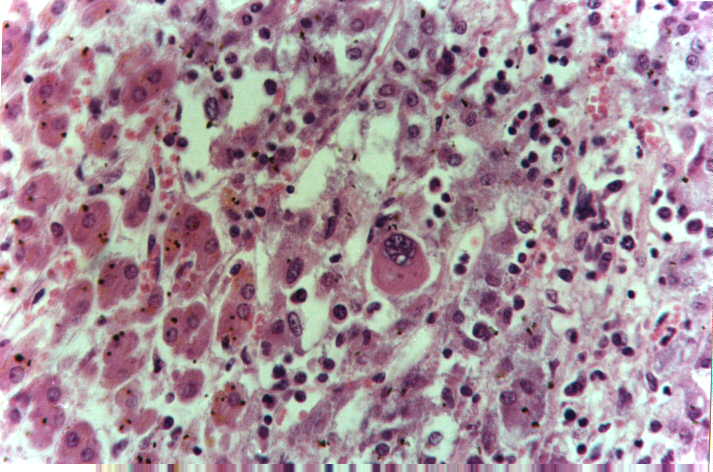 |
Right adrenal. There is only foci of extramedullary hematopoiesis, with no fat cells associated and without compression of glandular cells. The trilineage hematopoietic cells are between medullary and cortical cells. There are megakaryocyte (center), erithrocyte and myeloid lineages in all stages of differentiation, all of them typical. HE, 40x. |
|
|