| Paper # 095 | Versión
en Español |
| Paper # 095 | Versión
en Español |
Andréa Rodrigues Cordovil Pires, Luciana Wernersbach Pinto.
[Title] [Introduction] [Materials and Methods] [Results] [Discussion] [Bibliography]
|
|
|
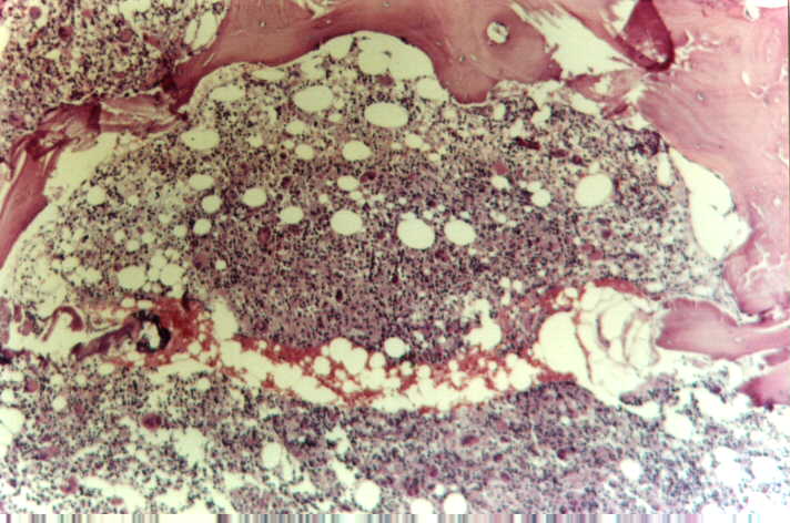
Bone marrow tissue from biopsy specimen. It shows marked cellularity, with a moderate increase in megakaryocyte number, many of them atypical.HE, 40x. |
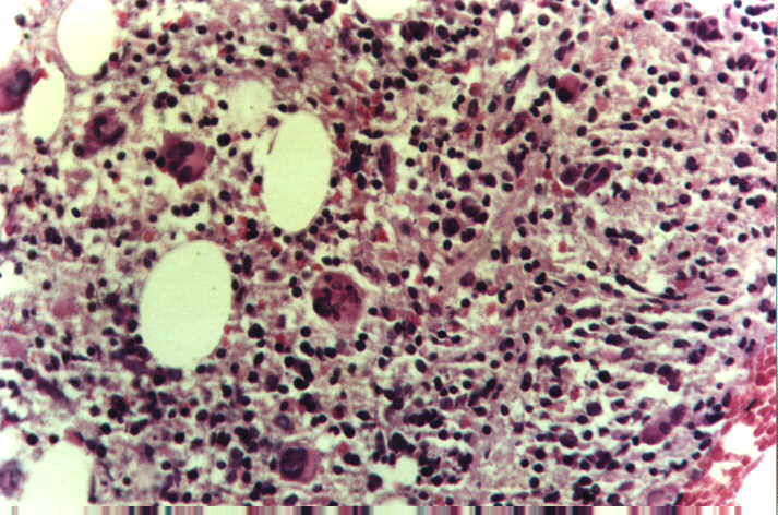 |
Bone marrow tissue from biopsy specimen. It shows marked cellularity, with a moderate increase in megakaryocyte number, many of them atypical, hyperlobulated and hyperchromatic.There is also a hyperplastic left shift of the myeloid lineage and some increase in connective tissue. HE, 40x. |
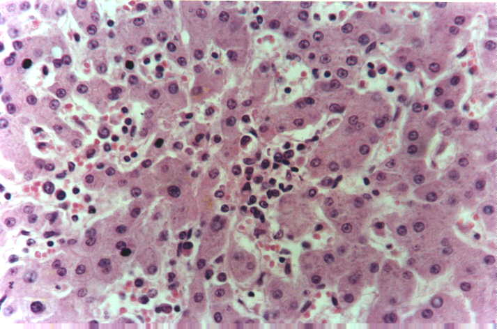 |
Liver tissue from autopsy. It shows hematopoietic islands inside sinusoids, predominantly with myeloid and erithroid lineages. There are also scattered megakaryocytes in other areas not shown.There is no fibrosis. HE, 40x. |
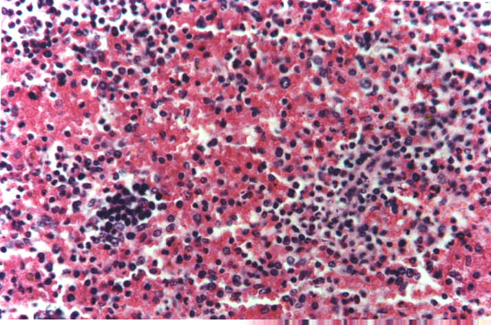 |
Spleen tissue from autopsy. It shows hematopoietic islands in red pulp, predominantly with myeloid and erithroid lineages. There are also scattered megakaryocytes in other areas not shown.There is no fibrosis. HE, 40x. |
|
|