| Paper # 096 | Versión
en Español |
| Paper # 096 | Versión
en Español |
Thymic microenvironmental changes in human congenital syphilis
Eliene C. Fonseca, Mônica P. Almeida, Évlin H. Maia, Dora M. F. Menezes and Wilson Savino*
[Title] [Introduction] [Materials and Methods] [Results] [Discussion] [Pictures] [Bibliography]
|
|
|
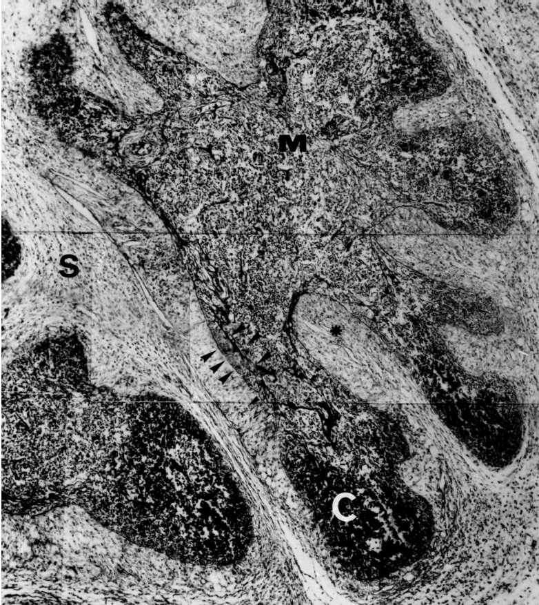
Congenital syphilis. Mounting of photomicrography showing reduction of the cortex (C), septal enlargement (S) with intralobular projetions (*). there is also basement membrane thickening and increase in reticular fibers inside the lobule (arrow heads), Paraffin section, Gomori's reticulin, 200x. |
 |
Congenital syphilis. Mounting of photomicrography showing lobular basement membrane thickening and intralobular "fibrosis" (arrows). Septa exibts intense plasmocitary infiltrate. Picrosirius not polarized, 100x. |
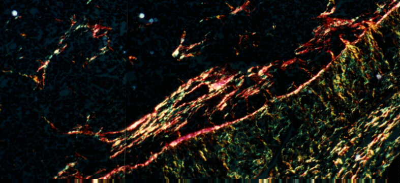 |
Congenital syphilis. Mounting of photomicrography showing lobular basement membrane thickening and intralobular "fibrosis", characterized by abundant type I collagen (arrows). Septa exibts intense plasmocitary infiltrate, area with predominance of type III collagen near basement membrane (perpendicular fibers) and an area away from basement membrane characteristic of type I collagen, also containig type III collagen. Picrosirius, polarized, 100 x. |
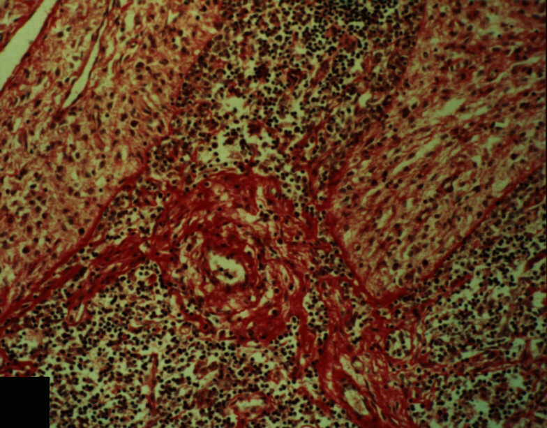 |
Congenital syphilis. Perivascular fibrosis and septa projections, associated with cortical lymphocyte depletion.Picrosirius, not polarized, 100 x. |
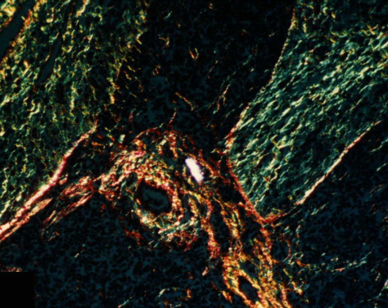 |
Congenital syphilis. Perivascular fibrosis containig type I collagen, basement membrane thickening and septa projections, associated with cortical lymphocyte depletion.Picrosirius, polarized, 100 x. |
|
|