| Paper # 001 | Versión en Español |
Marcial Garcia-Rojo, Jesús González, Ana
Morillo, Jesus Martin
[Title] [Introduction] [Materials and Methods] [Results] [Picture 4] [Discussion] [Bibliography]
|
|
Lymphoid cells were easily distinguishable from adenocarcinoma cells with the common leukocyte antigen staining (LCA x 400). |
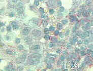
Adenocarcinoma signet ring cells were negative for all lymphoid markers. Lymphoid neoplastic cells were positive with pan-B cell markers like CD-20. (L26 x 630). |
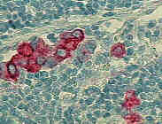
Malignant adenocarcinoma cells were identifiable by their positivity with epithelial markers. They showed an intense staining with the Common Epithelial Antigen, in contrast with the total absence of this marker in lymphoid cells (CEA x 400). |
Small cells of adenocarcinoma cells metastatic to lymph nodes were observed with low weight cytokeratins markers (Cytokeratins 8,18 &19 x 400). |
|