| Paper # 001 | Versión en Español |
Marcial Garcia-Rojo, Jesús González, Ana
Morillo, Jesús Martín
[Title] [Introduction] [Materials and Methods] [Results] [Picture 3] [Picture 5] [Discussion] [Bibliography]
|
|
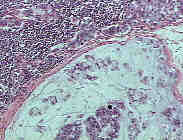
All the lymph nodes examined showed an effacement of the architecture due to a malignant lymphoma. Furthermore, marginal sinus of this node and adjacent perinodal adipose tissue were infiltrated by metastatic adenocarcinoma cell with abundant extracellular mucin (H&E x 40). |
Metastatic adenocarcinoma cells had the same signet ring appearance as the gastric tumor. In the lymph node they were located mainly in marginal sinus. The rest of the node was occupied by a low grade lymphoma type immunocytoma. (H&E x 100). |
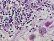
Neoplastic lymphocytes in the lymph node showed the same plasmacytoid characteristics as the lymphoma diagnosed six months earlier. Signet ring adenocarcinoma cells infiltrated extensively the perinodal soft tissues (PAS x 400). |
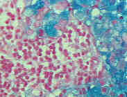
Adenocarcinoma cells metastatic to the lymph nodes were very easily identifiable with the mucin stains (Colloidal Iron x 400). |
|