| Paper # 001 | Versión en Español |
Marcial Garcia-Rojo, Jesús González, Ana
Morillo, Jesus Martin
[Title] [Introduction] [Materials and Methods] [Results] [Picture 1] [Picture 3] [Discussion] [Bibliography]
|
|
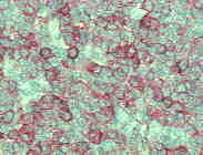
Tumoral cells with characteristic lymphoplasmacytoid nuclear and cytoplasmic features showed an intense stainning with pan-B cell markers like CD-45A (L26 x 400). |
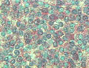
Non tumoral small lymphocytes were frequently observed. They were demonstrated with pan-T cell markers like CD45RO (UCHL-1 x 400). |
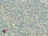
Neoplastic lymphocytes showed monoclonality in light chain immunoglobulin production. Only stainning for Kappa light chain was achieved (Kappa light chain x 630). |
Only scarce mature plasma cells were found to be positive with Lambda light chain. Note a tumoral cell in mitosis in upper right corner (Lambda light chain x 400). |
|
|